
- Title
-
Plate-like Guanine Biocrystals Form via Templated Nucleation of Crystal Leaflets on Preassembled Scaffolds
- Authors
- Eyal, Z., Deis, R., Varsano, N., Dezorella, N., Rechav, K., Houben, L., Gur, D.
- Source
- Full text @ J. Am. Chem. Soc.

Synthetic and biogenic guanine crystals have distinct morphologies. (A) A synthetic guanine crystal with a prismatic morphology, where the (100) crystallographic plane is the fastest growing direction. (B) A biogenic plate-like guanine crystal in a zebrafish eye. (C) Schematic illustration of both synthetic and biogenic crystals, where the (100) crystallographic plane is constructed from H-bonded molecular layers. (D) A zebrafish larva at 5 days post-fertilization contains guanine crystals in its eyes and skin. Insets show higher magnifications of the eye (cyan) and crystal-containing iridophores (pink). A and B are SEM micrographs and D shows incident light images. |

Early iridosomes contain several crystal leaflets that later coalesce into a single plate-like crystal. (A) An illustration of the zebrafish larva eye showing the iridophore (blue) and melanophore layers (purple), next to the lens. The anatomical location of the sections and areas that were taken for cryo-SEM (B-D) and cryo-FIB-SEM (E-G) are marked by a black rectangle and a black box, respectively. (B-D) Cryo-SEM images of freeze-fractured surfaces of a zebrafish larva eye and the iridophores within it. (B) A low magnification of the eye surface. Iridophore and melanophore layers are pseudocolored in blue and purple, respectively. (C) A close-up view of an iridophore next to a melanophore. (D) Iridosomes at different maturation stages. (E-G) Cryo-FIB-SEM images and 3D representations of segmented milled sections from a freeze-fractured zebrafish larva eye. (E) An illustration showing the stack of images of consecutive volumetric sections, with pseudocolored iridophore and melanophore layers. (F) An eye iridophore 3D representation of the area marked by a dashed rectangle in E. Mature elongated iridosomes and round developing iridosomes are marked in blue, nucleus is marked in gray. The early 200–300-nm-long iridosomes shown in (Gi-Giii) are marked with white arrows. (Gi-Giii) Cryo-FIB-SEM micrographs of different planes within the iridophore shown in (F). White arrows mark the iridosomes whose 3D reconstructions are shown in the insets. |
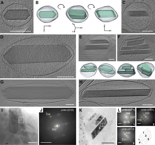
High-resolution cryo-TEM shows the morphological sequence of (100) crystal plates within iridosomes. (A, C–H) CryoET reconstructions of iridophores, isolated from zebrafish larvae at different developmental stages, show crystals either face-on (A,D,G) or edge-on (C,E,F,H) with respect to the electron beam. (B) A 3D representation of a crystal-containing iridosome from different angles. Bottom panels in (E) and (F) show 3D representations of leaflet-containing iridosomes. (I,K) Cryo-4D-STEM bright field images of iridosomes with face-on (I) or edge-on (K) orientated crystals. (J) Electron diffractions taken from the area marked by a white rectangle in (I). (L) Electron diffractions taken from the area marked by a white rectangle in (K), and an illustration showing the orientation of the a axes of the crystals (bottom right corner). A-I, K, scale bars are 100 nm. J, L, scale bars are 5 nm–1. |

Biogenic plate-like guanine crystals form via templated nucleation of (100) crystal leaflets on preassembled protein fibers. (A-E) CryoET reconstructions of iridophores isolated from zebrafish larvae at different maturation stages. Scale bars are 100 nm. (A) An early iridosome showing a scaffold of parallel protein fibers. (B) A more developed iridosome, in which the initial nucleation of (100) crystal leaflet on a protein scaffold is taking place. (C) Several individual leaflets, imaged edge-on, with their (100) crystal plane parallel to each other. (D) The leaflets have almost completely coalesced into a single crystal. (E) A mature iridosome, where the leaflets have completely merged into a single crystal. (F) Schematic illustration of the proposed crystallization mechanism. |