
Fig. 3
- ID
- ZDB-FIG-230515-3
- Publication
- Eyal et al., 2022 - Plate-like Guanine Biocrystals Form via Templated Nucleation of Crystal Leaflets on Preassembled Scaffolds
- Other Figures
- All Figure Page
- Back to All Figure Page
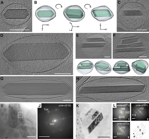
High-resolution cryo-TEM shows the morphological sequence of (100) crystal plates within iridosomes. (A, C–H) CryoET reconstructions of iridophores, isolated from zebrafish larvae at different developmental stages, show crystals either face-on (A,D,G) or edge-on (C,E,F,H) with respect to the electron beam. (B) A 3D representation of a crystal-containing iridosome from different angles. Bottom panels in (E) and (F) show 3D representations of leaflet-containing iridosomes. (I,K) Cryo-4D-STEM bright field images of iridosomes with face-on (I) or edge-on (K) orientated crystals. (J) Electron diffractions taken from the area marked by a white rectangle in (I). (L) Electron diffractions taken from the area marked by a white rectangle in (K), and an illustration showing the orientation of the a axes of the crystals (bottom right corner). A-I, K, scale bars are 100 nm. J, L, scale bars are 5 nm–1. |