
- Title
-
WNK1-OSR1 Signaling Regulates Angiogenesis-Mediated Metastasis towards Developing a Combinatorial Anti-Cancer Strategy
- Authors
- Hou, C.Y., Ma, C.Y., Lin, Y.J., Huang, C.L., Wang, H.D., Yuh, C.H.
- Source
- Full text @ Int. J. Mol. Sci.
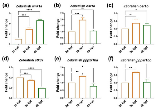
During embryonic angiogenesis, wnk1a and osr1b are upregulated. The expression of wnk1 and its downstream effectors during embryonic angiogenesis. (a) wnk1a, (b) osr1a, (c) osr1b, (d) stk39, (e) ppp2r1ba and (f) ppp2r1bb in zebrafish embryos stages from 24 h post-fertilization (hpf) to 48 hpf during embryonic angiogenesis. For each experiment, we performed three replicates, and for each qPCR, there were triplicates for each sample. Thirty larvae were included in each time point. One-way ANOVA analysis followed by multiple analysis was used to identify the statistical significance between groups; *: p ≦ 0.05; **: p ≦ 0.01; ***: p ≦ 0.001; ****: p ≦ 0.0001; ns: no-significance. |

Tumor-induced angiogenesis promotes hepatoma cell proliferation and migration. (a) The representative stacked images of 1 day post injection (dpi) and 3 dpi after red fluorescence-labeled Hep3B cells were injected into green fluorescence blood vessel embryos of Tg(fli1:EGFP)-control, Tg(fli1:EGFP)xTg(fli1:wnk1a)-WNK1(OE), and Tg(fli1:EGFP)xTg(fli1:CreERT2;myl7:EGFP)xTg(loxP-wnk1a-DsRed-loxP) after RU486 treated for knockout wnk1a-(WNK1(KO)). The images showed the tumor-induced angiogenesis with ectopic vessels surrounding the Hep3B cells. Quantification of area change comparing 3 dpi versus 1 dpi in control, WNK1 OE, or WNK1 KO embryos. (b) The representative images for control, WNK1 OE, or WNK1 KO embryos, red arrows indicate the migrated cells in WNK1(OE). Quantification of migration behavior comparing 3 dpi versus 1 dpi after red fluorescence-labeled Hep3B cells were injected into control, WNK1(OE), and WNK1(KO) embryos. We performed three replicates, and ten to thirty embryos were included in each experiment. One-way ANOVA analysis followed by multiple analysis was used to identify the statistical significance between groups; *: p ≦ 0.05; **: p ≦ 0.01; ****: p ≦ 0.0001; ns: no-significance. The scale bar is 500 µm in length. |
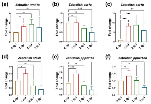
The expressions of wnk1a and osr1b are upregulated in endothelial cells during tumor-induced angiogenesis. The expression of wnk1a and its downstream effectors during tumor-induced angiogenesis. (a) wnk1a, (b) osr1a, (c) osr1b, (d) stk39, (e) ppp2r1ba and (f) ppp2r1bb in zebrafish embryos from 0 dpi to 3 dpi during tumor-induced angiogenesis. We performed three to four replicates, and for each qPCR, there were triplicates for each sample. Thirty embryos were included in each experiment. One-way ANOVA analysis followed by multiple analysis was used to identify the statistical significance between groups. *: p ≦ 0.05; **: p ≦ 0.01; ***: p ≦ 0.001; ns: no-significance. |
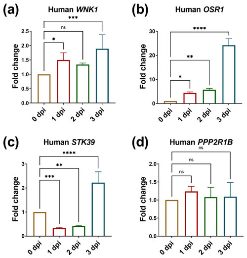
The expression of WNK1 and OSR1 is upregulated in hepatoma cells during tumor-induced angiogenesis promoting hepatoma cell proliferation. The expression of WNK1 and its downstream effectors in hepatoma cells during tumor induced angiogenesis. (a) WNK1, (b) OSR1, (c) STK39, (d) PPP2R1B in zebrafish embryos carrying hepatoma cells from 0 dpi to 3 dpi during tumor-induced angiogenesis. We performed three to four replicates, and for each qPCR, there were triplicates for each sample. Thirty embryos were included in each experiment. One-way ANOVA analysis followed by multiple analysis was used to identify the statistical significance between groups. *: p ≦ 0.05; **: p ≦ 0.01; ***: p ≦ 0.001; ****: p ≦ 0.0001; ns: no-significance. |
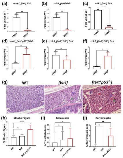
The expression of cell proliferation markers and histopathological features from H&E stain in [tert] and [tert x p53−/−] transgenic fish. The expression of proliferation marker in [tert] fish at 15 dpf and 30 dpf (a) ccne1, (b) cdk1, and (c) cdk2, and in [tert x p53−/−] fish at 15 dpf and 30 dpf (d) ccne1, (e) cdk1, and (f) cdk2. *: p ≦ 0.05; **: p ≦ 0.01; ***: p ≦ 0.001. (g) The representative images of H&E stain of WT, [tert], and [tert x p53−/−] fish at 30 dpf. The blue circle indicates the mitotic features, the green square points out the trinucleated hepatocytes, the red square denotes the karyomegalic cells. The scale bar is 20 µm in length. The quantification of histopathological features from H&E stain: (h) mitotic figures, (i) trinucleated hepatocytes, (j) karyomegalic cells are increased in WT, [tert], and [tert x p53−/−] fish at 30 dpf. We performed three replicates, and for each qPCR, there were triplicates for each sample. Ten embryos were included in each experiment. One-way ANOVA analysis followed by multiple analysis was used to identify the statistical significance between groups. *: p ≦ 0.05; **: p ≦ 0.01; ***: p ≦ 0.001; ****: p ≦ 0.0001; ns: no-significance. EXPRESSION / LABELING:
PHENOTYPE:
|

Upregulated wnk1a, stk39, and osr1b, and downregulated ppp2r1ba and ppp2r1bb in 15 dpf [tert] and [tert x p53−/−] transgenic fish. The expression of wnk1a and its downstream effectors in WT, [tert], and [tert x p53−/−] fish at 15 dpf. (a) wnk1a, (b) osr1a, (c) osr1b, (d) stk39, (e) ppp2r1ba, and (f) ppp2r1bb in 15 dpf [tert] and [tert x p53−/−] transgenic fish compared to WT. We performed three replicates, and for each qPCR, there were triplicates for each sample. Ten larvae were included in each experiment. One-way ANOVA analysis followed by multiple analysis was used to identify the statistical significance between groups. *: p ≦ 0.05; **: p ≦ 0.01; ****: p ≦ 0.0001; ns: no-significance. EXPRESSION / LABELING:
PHENOTYPE:
|
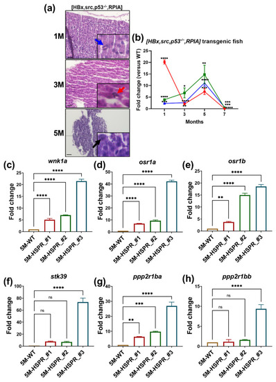
H&E stain and QPCR analysis reveal that [HBx,src,p53−/−,RPIA] transgenic fish developed HCC at 5 months of age. (a) The representative images of H&E staining of 1, 3, 5M [HBx,src,p53−/−,RPIA] transgenic fish. The scale bar is 20 µm in length. (b) The expression of proliferation marker-ccne1, cdk1, and cdk2 of 1, 3, 5M [HBx,src,p53−/−,RPIA] transgenic fish. The expression of wnk1a and its downstream effectors in WT and [HBx,src,p53-/-,RPIA] transgenic fish at 5 months of age. (c) wnk1a, (d) osr1a, (e) osr1b, (f) stk39, (g) ppp2r1ba, and (h) ppp2r1bb at 5 months of #2, #3, #4 independent [HBx,src,p53−/−,RPIA] transgenic fish compared to WT. We performed three replicates, and for each qPCR, there were triplicates for each sample. Multiple adult fish were included in each experiment. One-way ANOVA analysis followed by multiple analysis was used to identify the statistical significance between groups. *: p ≦ 0.05; **: p ≦ 0.01; ***: p ≦ 0.001; ****: p ≦ 0.0001; ns: no-significance. |

Knockdown of WNK1 in HUVEC cells significantly decreases the co-cultured hepatoma cell migration, and OSR1 overexpression can rescue the reduced cell migration caused by shWNK1 knockdown in endothelial cells. (a) The protein levels of WNK1 and OSR1 with WNK1 knockdown and/or OSR1 overexpression in HUVEC cells were detected by Western blot. (b) Quantification of WNK1 Western blot indicated that knockdown WNK1 (shWNK1-1 and shWNK1-2) in HUVEC cells significantly decreases the WNK1 protein levels. (c) Quantification of OSR1 Western blot indicated that overexpression of OSR1 in HUVEC cells significantly increases the OSR1 protein level, knockdown WNK1 (shWNK1-1 and shWNK1-2) in HUVEC cells significantly decreases the OSR1 protein levels. (d) HUVEC cells modulated the expression of WNK1 and OSR1 and were seeded in the lower chamber while HepG2 hepatoma cells were loaded in the upper chamber of the transwell plate. (e) Representative images of hepatoma cell migration. The scale bar is 200 µm in length. (f) Quantified data of the transwell migration assay shows that HUVEC cells with WNK1 knockdown significantly decrease the migrated hepatoma cells, indicating that WNK1 plays an important role in endothelial cells for stimulating hepatoma cell migration. Overexpression of OSR1 in shWNK1 knockdown HUVEC cells can revert the reduced hepatoma cell migration caused by shWNK1, indicating that OSR1 is downstream of endothelial WNK1 promoting hepatoma cell migration. We performed three replicates for each Western blot and transwell analysis. One-way ANOVA analysis followed by multiple analysis of variance was used to identify the statistical significance between groups. *: p ≦ 0.05; **: p ≦ 0.01; ***: p ≦ 0.001; ****: p ≦ 0.0001; ns: no-significance. |

Overexpression of WNK1 in HUVEC cells significantly increases the migration of HepG2 cells. (a) The protein levels of WNK1 were examined by Western blot. (b) Quantification of Western blot indicates that overexpression of WNK1 in HUVEC cells significantly increases the WNK1 protein levels. (c) HUVEC cells with WNK1 overexpression were seeded in the lower chamber while HepG2 hepatoma cells were loaded in the upper chamber of the transwell plate. (d) Representative images of hepatoma cell migration. The scale bar is 200 µm in length. (e) Quantified data of the transwell migration assay shows that WNK1 overexpression in HUVEC cells significantly increases the migrated HepG2 cells, indicating that WNK1 plays an important role in endothelial cells for stimulating hepatoma cell migration. We performed three replicates for each Western blot and transwell analysis. Unpaired Student’s t-test (two tailed test) was used for statistical analysis. *: p ≦ 0.05; **: p ≦ 0.01. |

Overexpression of WNK1 in HepG2 cells significantly increases the migrated cells, and the co-expression of PPP2R1A can decrease the migrated cells caused by WNK1 overexpression in hepatoma cells. (a) The protein levels of WNK1 and PPP2R1A were examined by Western blot. (b) Western blot indicates that overexpression of WNK1 in HepG2 cells significantly increases the WNK1 protein levels. (c) Overexpression of PPP2R1A in HepG2 cells significantly increases the PPP2R1A protein levels. (d) The modulated expression of WNK1 and PPP2R1A in HepG2 hepatoma cells that were seeded in the upper chamber while HUVEC cells were loaded in the lower chamber of the transwell plate. (e) Representative images of hepatoma cell migration. The scale bar is 200 µm in length. (f) Quantified data of the transwell migration assay reveal that overexpression of WNK1 in HepG2 cells significantly increases the migrated cells, indicating that WNK1 plays an important role in hepatoma cells for stimulating hepatoma cell migration. Overexpression of PPP2R1A in HepG2 cells can revert the increased migrated cells caused by WNK1, indicating that PPP2R1A is a downstream effector for hepatoma cell migration. We performed three replicates for each Western blot and transwell analysis. One-way ANOVA analysis followed by multiple analysis of variance was used to identify the statistical significance between groups. *: p ≦ 0.05. |
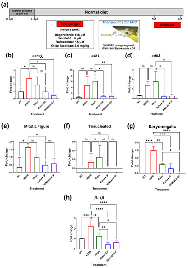
WNK1–OSR1 axis inhibitors combined with oligo-fucoidan attenuate HCC proliferation. (a) The treatment scheme of WNK1 and OSR1 inhibitors combined with oligo-fucoidan in [HBx,src,p53−/−,RPIA] HCC transgenic fish. The expression of proliferation markers (b) ccne1, (c) cdk1, and (d) cdk2 in various combinations of drug-treated 5-month-old [HBx,src,p53−/−,RPIA] (HSPR) transgenic fish compared to WT. Rego: Regorafenib, Rafox: Rafoxanide as OSR1 inhibitor; WNK463 as WNK1 inhibitor; OF: oligo-fucoidan. The quantification of the histopathological features derives from H&E stain: (e) mitotic figures, (f) trinucleated hepatocytes, (g) karyomegalic cells in various combinations of drug-treated 5-month-old [HBx,src,p53−/−,RPIA] transgenic fish compared to WT. (h) The expression of cell senescence marker il1β in various combinations of drug-treated 5-month-old [HBx,src,p53−/−,RPIA] (HSPR) transgenic fish compared to WT. We performed three replicates, and for each qPCR, there are triplicates for each sample. Five adult fish are included in each experiment. One-way ANOVA analysis followed by multiple analysis of variance was used to identify the statistical significance between groups. *: p ≦ 0.05; **: p ≦ 0.01; ***: p ≦ 0.001; ****: p ≦ 0.0001; ns: no-significance. |
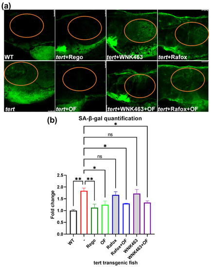
Oligo-fucoidan reduces the elevated senescence-associated β-galactosidase (SA-β-gal) expression by WNK1–OSR1 axis inhibitors in tert transgenic fish. We use Tg(fabp10a:tert; myl7:GFP) transgenic fish to further verify whether drugs can prevent cellular senescence. These fish were fed with a normal diet from 5 dpf to 10 dpf old and treated with several drugs: Regorafenib (0.5 μM), oligo-fucoidan (0.3 mg/mL), WNK463 (0.625 μM) with or without oligo-fucoidan, Rafoxanide(0.3 μM) with or without oligo-fucoidan, and were then fixed and stained on 11 dpf. (a) The SA-β-gal expression in various combinations of drug-treated tert transgenic fish after various drug treatments. WT: AB(WT) fish as control, tert: Tg(fabp10a:tert; myl7:GFP) fish; OF: oligo-fucoidan-treated tert transgenic fish; WNK463: WNK1 inhibitor WNK463-treated tert transgenic fish; WNK463+OF: WNK463 plus oligo-fucoidan-treated tert transgenic fish; Rafox: OSR1 inhibitor Rafoxanide-treated tert transgenic fish; Rafox+OF: Rafoxanide plus oligo-fucoidan-treated tert transgenic fish. The scale bar is 100 µm in length. The orange circle indicates the location of the liver. (b) Quantification of SA-β-gal staining. For each experiment, we performed three replicates. Ten larvae were included in each experiment. One-way ANOVA analysis followed by multiple analysis of variance was used to identify the statistical significance between groups. *: p ≦ 0.05; **: p ≦ 0.01; ns: no-significance. PHENOTYPE:
|