
Figure 7
|
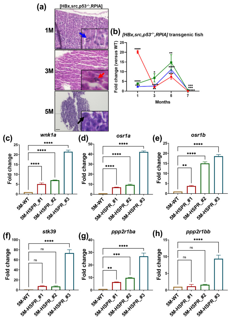
Figure 7
H&E stain and QPCR analysis reveal that [HBx,src,p53−/−,RPIA] transgenic fish developed HCC at 5 months of age. (a) The representative images of H&E staining of 1, 3, 5M [HBx,src,p53−/−,RPIA] transgenic fish. The scale bar is 20 µm in length. (b) The expression of proliferation marker-ccne1, cdk1, and cdk2 of 1, 3, 5M [HBx,src,p53−/−,RPIA] transgenic fish. The expression of wnk1a and its downstream effectors in WT and [HBx,src,p53-/-,RPIA] transgenic fish at 5 months of age. (c) wnk1a, (d) osr1a, (e) osr1b, (f) stk39, (g) ppp2r1ba, and (h) ppp2r1bb at 5 months of #2, #3, #4 independent [HBx,src,p53−/−,RPIA] transgenic fish compared to WT. We performed three replicates, and for each qPCR, there were triplicates for each sample. Multiple adult fish were included in each experiment. One-way ANOVA analysis followed by multiple analysis was used to identify the statistical significance between groups. *: p ≦ 0.05; **: p ≦ 0.01; ***: p ≦ 0.001; ****: p ≦ 0.0001; ns: no-significance.