
- Title
-
Zebrafish: an important model for understanding scoliosis
- Authors
- Xie, H., Li, M., Kang, Y., Zhang, J., Zhao, C.
- Source
- Full text @ Cell. Mol. Life Sci.
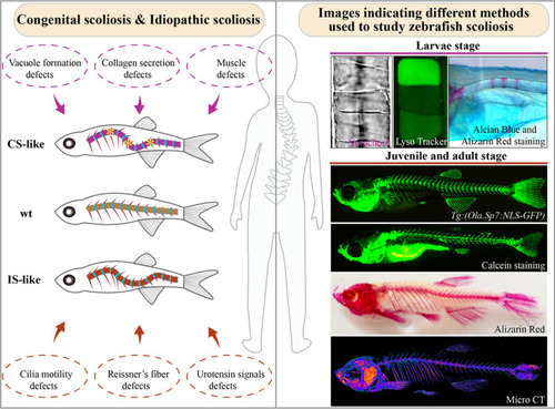
Zebrafish as a vertebrate model for scoliosis. Left: Different types of scoliosis and their potential causes from zebrafish studies. Scoliosis due to neuromuscular defects is also illustrated as CS like group. Asterisks indicate the abnormally developed vertebrae in CS-like zebrafish mutants. Right: Various bone staining and imaging methods used to evaluate skeleton development in zebrafish. At larvae stages, the notochord and vacuoles can be easily visualized via bright-field image (top left) or LysoTracker dye staining (top middle). Skeletal development in zebrafish can be visualized via alcian blue-Alizarin red double staining (top right). At juvenile or adult stages, skeleton development can be visualized via transgenic labeling, calcein staining, Alizarin red staining or micro-CT |
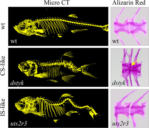
Phenotypes of CS-like and IS-like zebrafish mutants. Left, Micro-CT images showing the morphology of spine in wild type, |
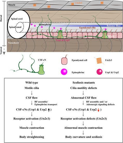
Model illustrating the Urotensin signaling pathway during body axis development of zebrafish. The beating of ependymal motile cilia drives CSF flow, which mediates the transmission of epinephrine signals and the assembly of Reissner’s fiber. The adrenergic signals activate the expression of Urotensin neuropeptides in the CSF-cNs (Urp1 and Urp2). Finally, the urotensin neuropeptides may further activate the Uts2r3 receptor localized to the dorsal muscle fibers and promote body straightening. Abnormalities in the Urotensin signaling pathway will induce either body curvature in larvae or scoliosis in adult zebrafish |