FIGURE SUMMARY
Title

Cholenic acid derivative UniPR1331 impairs tumor angiogenesis via blockade of VEGF/VEGFR2 in addition to Eph/ephrin

Authors
Rusnati, M., Paiardi, G., Tobia, C., Urbinati, C., Lodola, A., D'Ursi, P., Corrado, M., Castelli, R., Wade, R.C., Tognolini, M., Chiodelli, P.
Source
Full text @ Cancer Gene Ther.

Binding of UniPR1331 to VEGFR2.

a Chemical structure of UniPR1331. b SPR sensorgrams showing the binding of UniPR1331 (40 μM) to the VEGFR2-Fc-coated or to the control Fc-coated surfaces. c Blank-subtracted SPR sensorgrams derived from injection of UniPR1331 on the VEGFR2-Fc surface. d Steady-state analysis obtained by Scatchard’s plot analysis of the bound RU values at equilibrium from b. White dot represents UniPR1331 binding to a control VEGF-coated surface. e ELISA-based competition experiments: inhibition curves of the binding of biotinylated ephrin-A1-Fc or VEGF to immobilized EphA2-Fc and VEGFR2 ectodomain by UniPR1331. Data in bd are representative of other three experiments that gave similar results. Data in e are expressed as percent of binding in respect to control without inhibitor and are the mean ∓ S.E.M. of 3–6 independent experiments. f Modeled structure of the UniPR1331/VEGFR2 complex from the final frames of MD showing the key residues involved in the interaction. D2–D3 domain of VEGFR2 is depicted in white cartoons representations. VEGFR2 residues involved in the interaction and UniPR1331 are shown in gray and magenta sticks (oxygen in red, nitrogen in blue). Hydrophilic (H-bonds and salt links) and hydrophobic interactions are indicated with yellow dashed and green dotted lines.

Effect of UniPR1331 on VEGF/VEGFR2 binding at the cell surface.

a Chlorate-treated ECD-VEGFR2-EYFP GM7373 cells were incubated with VEGF and UniPR1331 and analyzed in immunofluorescence with anti-VEGF antibody. Fluorescence was quantified using ImageJ software as corrected total cell fluorescence: integrated density − (area of selected cell × mean fluorescence of background). Data are the mean ± S.E.M. of measurements on 25–35 cells for each sample from two independent experiments. (*P < 0.0001 in respect to cell treated with VEGF alone). b Microphotographs of ECD-VEGFR2-YFP GM7373 incubated with VEGF and UniPR1331. Scale bar: 10 μM. c ECD-VEGFR2-YFP A745 CHO cells were added to HSPG-bearing CHO-K1 monolayers with VEGF and UniPR1331. Adherent cells were photographed and counted after 2 h of incubation. Data are the mean ± S.E.M. of cell count in six microscopic fields. d Microphotographs of ECD-VEGFR2-YFP A745 CHO-K1 incubated on HSPG-bearing CHO-K1 monolayers with VEGF and UniPR1331.

Effect of UniPR1331 on VEGFR2 activation.

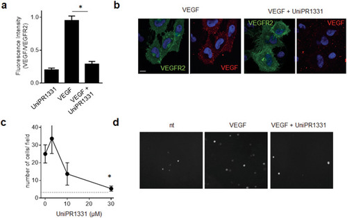
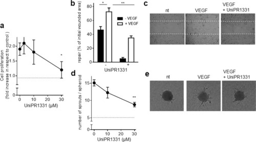
The indicated cells were left unstimulated or stimulated with 10 ng/ml VEGF (a, b) or 30 ng/ml FGF2 (c) with UniPR1331 (30 μM in ac or increasing concentrations in b). Then, cells were analyzed by WB with anti-P-VEGFR2 (a, b) or anti-P-FGFR1 (c) antibody. Uniform loading was confirmed with anti-VEGFR-2 or anti-tubulin antibody. The results shown are representative of other two that gave similar results. The single lanes have been cropped and reorganized for an easy comparison. b Densitometric quantification of P-VEGFR2 immunoreactive bands normalized to the expression levels of VEGFR2. (mean ± S.E.M. of three independent experiments, *P < 0.005).

Effect of UniPR1331 on VEGFR2 internalization and signal transduction.

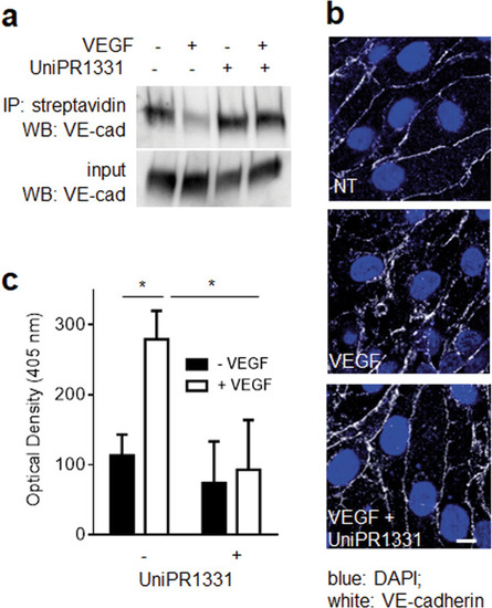
HUVECs unstimulated or stimulated with VEGF and UniPR1331 were analyzed for VEGFR2 internalization by WB (a) or by FACs analysis (b) or for the activation of VEGF-dependent signal transduction by WB with anti-P-ERK1/2 (c) or anti-P-PLC-γ (d) antibody. Uniform loading was confirmed by WB with anti-FAK (c) or anti-tubulin (d) antibody. The results shown are representative of two to three other experiments that gave similar results.

Effect of UniPR1331 on VEGF-induced VE-cadherin internalization and permeabilization in ECs.

a HUVECs unstimulated or stimulated with VEGF and UniPR1331 were analyzed for VE-cadherin internalization by WB. b Microphotographs of GM7373-VEGFR2 cells incubated with VEGF and UniPR1331 and immunostained for VE-cadherin (scale bar: 10 μm). c Monolayer of GM7373-VEGFR2 cells unstimulated or stimulated with VEGF and UniPR1331 were evaluated for permeability as described in “Material and methods.” The results shown are the mean ± S.E.M. of three independent experiments; *P < 0.05.

Effect of UniPR1331 on VEGF/VEGFR2-dependent EC proangiogenic activation.

a HUVECs stimulated with VEGF and increasing concentrations of UniPR1331 were counted using the MACSQuant Analyzer. Data are expressed as proliferation fold increase in respect to HUVECs cells left untreated (dashed line). b HUVEC monolayers were wounded and incubated with VEGF and UniPR1331. Then, the extension of the repaired wound area was evaluated. Data are expressed as percent or repaired area in respect to the extension of the original wound. c Microphotographs of wounded HUVEC monolayers taken after 24 h of incubation with or without VEGF and UniPR1331. Dashed lines mark the edge of the wound at t0. d HUVEC spheroids embedded in fibrin gel were incubated with VEGF and UniPR1331. Then, radially growing cell sprouts were counted. e Microphotographs of spheroids incubated with VEGF and UniPR1331. Data shown in a, b, and d are the mean ± S.E.M. of three independent experiments (*P < 0.05 and **P < 0.01).

Effect of UniPR1331 on in vivo angiogenesis.

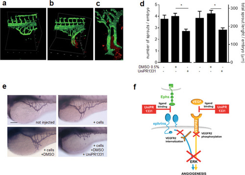
3D images reconstruction of SIV in Tg(fli1:EGFP)y1 zebrafish embryos in the absence (a) or in the presence (b) of U251 cells (in red). c High magnification of the image reconstruction of b showing the detail of ectopic SIV vessels converging toward U251 cells. d Evaluation of the angiogenic response: quantification of AP + ectopic sprouts are expressed as number of sprouts/embryo and cumulative length/embryo normalized in respect to noninjected embryos (number of embryos analyzed: U251 alone, 32; U251 + DMSO, 51; U251 + UniPR1331, 47). Data are the mean ± S.E.M. of three independent experiments (*P < 0.05). e Representative images of the angiogenic response used for the quantifications reported in d. Scale bar: 100 µm. f Schematic representation of the multitarget mechanisms of action of UniPR1331. By binding to Ephs, UniPR1331 inhibits VEGFR2 internalization required for downstream signaling. Simultaneously, UniPR1331 binds VEGFR2, hampering its interaction with VEGF and receptor phosphorylation. Both these inhibitions contribute to prevent phosphorylation of ERK1/2, a key second messenger for EC proangiogenic activation.

Acknowledgments
This image is the copyrighted work of the attributed author or publisher, and ZFIN has permission only to display this image to its users. Additional permissions should be obtained from the applicable author or publisher of the image. Full text @ Cancer Gene Ther.