IMAGE

Fig. 2

ID
ZDB-IMAGE-220722-3
Source
Figures for Rusnati et al., 2021
Image
Figure Caption

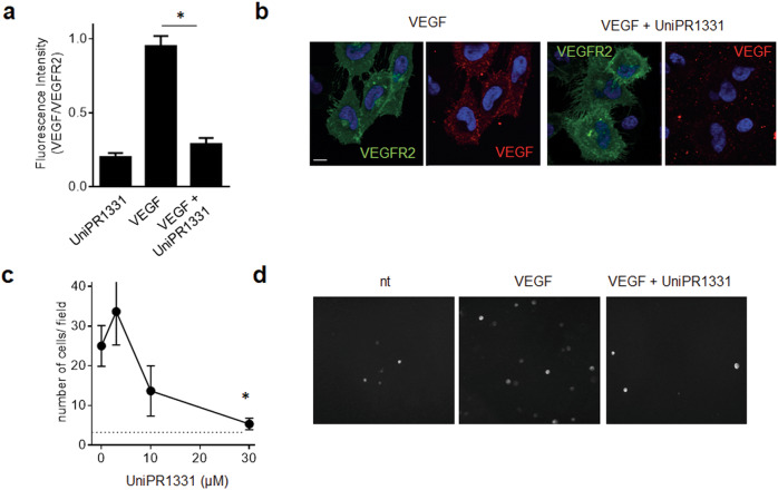
Fig. 2 Effect of UniPR1331 on VEGF/VEGFR2 binding at the cell surface.

a Chlorate-treated ECD-VEGFR2-EYFP GM7373 cells were incubated with VEGF and UniPR1331 and analyzed in immunofluorescence with anti-VEGF antibody. Fluorescence was quantified using ImageJ software as corrected total cell fluorescence: integrated density − (area of selected cell × mean fluorescence of background). Data are the mean ± S.E.M. of measurements on 25–35 cells for each sample from two independent experiments. (*P < 0.0001 in respect to cell treated with VEGF alone). b Microphotographs of ECD-VEGFR2-YFP GM7373 incubated with VEGF and UniPR1331. Scale bar: 10 μM. c ECD-VEGFR2-YFP A745 CHO cells were added to HSPG-bearing CHO-K1 monolayers with VEGF and UniPR1331. Adherent cells were photographed and counted after 2 h of incubation. Data are the mean ± S.E.M. of cell count in six microscopic fields. d Microphotographs of ECD-VEGFR2-YFP A745 CHO-K1 incubated on HSPG-bearing CHO-K1 monolayers with VEGF and UniPR1331.

Acknowledgments
This image is the copyrighted work of the attributed author or publisher, and ZFIN has permission only to display this image to its users. Additional permissions should be obtained from the applicable author or publisher of the image. Full text @ Cancer Gene Ther.