IMAGE

Fig. 6

ID
ZDB-IMAGE-220722-7
Source
Figures for Rusnati et al., 2021
Image
Figure Caption

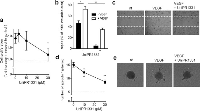
Fig. 6 Effect of UniPR1331 on VEGF/VEGFR2-dependent EC proangiogenic activation.

a HUVECs stimulated with VEGF and increasing concentrations of UniPR1331 were counted using the MACSQuant Analyzer. Data are expressed as proliferation fold increase in respect to HUVECs cells left untreated (dashed line). b HUVEC monolayers were wounded and incubated with VEGF and UniPR1331. Then, the extension of the repaired wound area was evaluated. Data are expressed as percent or repaired area in respect to the extension of the original wound. c Microphotographs of wounded HUVEC monolayers taken after 24 h of incubation with or without VEGF and UniPR1331. Dashed lines mark the edge of the wound at t0. d HUVEC spheroids embedded in fibrin gel were incubated with VEGF and UniPR1331. Then, radially growing cell sprouts were counted. e Microphotographs of spheroids incubated with VEGF and UniPR1331. Data shown in a, b, and d are the mean ± S.E.M. of three independent experiments (*P < 0.05 and **P < 0.01).

Acknowledgments
This image is the copyrighted work of the attributed author or publisher, and ZFIN has permission only to display this image to its users. Additional permissions should be obtained from the applicable author or publisher of the image. Full text @ Cancer Gene Ther.