FIGURE SUMMARY
Title

Identification of 3' UTR motifs required for mRNA localization to myelin sheaths in vivo

Authors
Yergert, K.M., Doll, C.A., O'Rouke, R., Hines, J.H., Appel, B.
Source
Full text @ PLoS Biol.

Sequence motifs derived from the <italic>mbpa</italic> 3′ UTR are sufficient for mRNA localization to myelin.

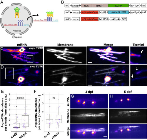
(A) Schematic representation of the MS2 mRNA reporter with motifs inserted upstream of the sv40 3′ UTR. Green box is motif 1, blue box is motif 2, and red box is motif 3. (B) Schematic representation of the primary sequences used to test the sufficiency of the motifs in (A). Green underline is motif 1, blue underline is motif 2, and red underline is motif 3. Top to bottom correspond to fmr1, eif4ebp2, and mbpa sequences. (C) Representative images of MS2 system after motifs from the mbpa, eif4ebp2, or fmr1 3′ UTRs were inserted into the MS2 mRNA reporter. Asterisks mark cell bodies. (D) Quantification of mRNA abundance in myelin sheaths from (A–C). (E) Quantification of mRNA abundance in myelin sheaths from individual insertions of each motif. Statistical analysis evaluated with Wilcoxon test. Scale bars, 10 μm. A minimum (n) of 6 embryos and 35 sheaths were used in each condition (D and E). The underlying numerical data can be found in S1 and S4 Data. 3' UTR, 3' untranslated region.

The <italic>mbpa</italic> 3′ UTR is sufficient to localize mRNA to myelin sheaths in living zebrafish.

(A) Schematic of the MS2 system to visualize mRNA localization in oligodendrocytes. sox10 regulatory DNA drives expression of nuclear-localized MS2 coat protein, NLS-MCP-EGFP (orange crescent and green star). mbpa regulatory elements drive expression of mRNA encoding mScarlet-CAAX fluorescent protein with a repetitive sequence that creates 24 stem loops (24xMBS). When co-expressed, the mRNA–protein complex is exported from the nucleus and localized via the 3′ UTR. (B) Schematic of MS2 expression plasmids used for transient expression in oligodendrocytes with target sequences for Tol2 transposase to facilitate transgene integration. (C and D) Representative images of localization directed by the mbpa (C) or control sv40 3′ UTR (D). Asterisks mark cell bodies with high expression levels of the nuclear-localized MCP-EGFP. Boxed areas are enlarged to highlight sheath termini (arrows). (E) Average mRNA abundance per myelin sheath, measured by EGFP fluorescence intensity normalized to the average intensity of the sv40 control. sv40: n = 5 larvae, 35 sheaths. mbpa: n = 6 larvae, 38 sheaths. (F) Average mRNA abundance per soma, measured by EGFP fluorescence intensity normalized to the average intensity of the sv40 control. sv40: n = 11 larvae, 20 cell bodies. mbpa: n = 15 larvae, 21 cell bodies. (G and H) Representative images of 2 myelinating oligodendrocytes expressing mRNA lacking the 24xMBS. NLS-MCP-EGFP remains in the nucleus at 3 dpf (G) and 5 dpf (H). Scale bars, 10 μm. Statistical significance evaluated using Wilcoxon test. The underlying numerical data can be found in S1 and S2 Data. 3' UTR, 3' untranslated region; dpf, days post fertilization.

Endogenous <italic>mbpa</italic> mRNA localizes to myelin sheaths between 3–5 dpf.

(A and B) Representative images of smFISH experiments using 4 dpf transgenic larva expressing EGFP-CAAX to mark oligodendrocytes. Images show sagittal sections of the hindbrain. DAPI stain labels nuclei. Sections were treated with smFISH probes designed to detect mbpa (A) or egfp (B) mRNA. Asterisks mark cell bodies and brackets mark myelin tracts. Scale bars, 10 μm. (C) Average mbpa mRNA density per cell body or equivalent volume of myelin from 3 to 5 dpf. Density was measured using the integrated density of fluorescence intensity in cell bodies and approximately equal volumes of myelin along the myelin tracts. A minimum (n) for each group was 3 larvae, 6 cell bodies, and 15 myelin regions. Statistical significance evaluated using Wilcoxon test. (D) Proportion of egfp or mbpa mRNA abundance in cell bodies compared to myelin tracts. A minimum (n) for each group was 3 larvae, 11 cell bodies, and 21 myelin regions. (E) Average mbpa mRNA density within individual sheaths plotted as a function of sheath length. Statistical significance evaluated using Spearman’s correlation coefficient. Shaded area represents 95% confidence interval. n = 7 embryos, 26 sheaths. The underlying numerical data can be found in S5S7 Data. dpf, days post fertilization; smFISH, single molecule fluorescent in situ hybridization.

The <italic>mbpa</italic> 3′ UTR is sufficient to localize mRNA to the leading edge of myelin sheaths during wrapping.

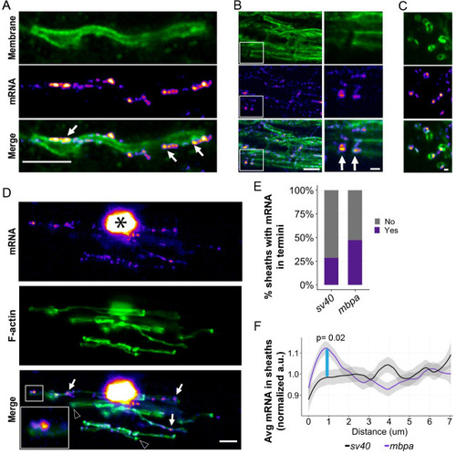
(A) smFISH images of a single optical section of a myelin sheath in a 3-dpf larva spinal cord. mbpa transcripts line the myelin sheath. Arrows highlight clusters of mbpa mRNA transcripts. (B) smFISH images of a single optical section of myelin tracts in the hindbrain of a 5-dpf larva. Boxed area magnified to highlight sheath termini (arrows). (C) smFISH images of a single optical section in transverse plane of myelin sheaths in a 5-dpf larva midbrain. Scale bars (A, B, and D), 5 μm; (C, boxed enlargements), 1 μm. (D) Representative images from MS2 system showing colocalization of mRNA containing mbpa 3′ UTR and F-actin in a myelinating oligodendrocyte. Asterisk marks the cell body, and boxes are magnified to highlight sheath termini. Arrows highlight sheaths with mRNA, and arrowheads highlight sheaths lacking mRNA. (E) Proportion of sheaths with mRNA enriched in sheath termini at 4 dpf using the MS2 system. Proportion measured as (sheaths with enrichment / number of sheaths) = 10/35 sv40, 18/38 mbpa. (F) Average fluorescence intensity of MS2 mRNA reporter containing the sv40 or mbpa 3′ UTRs across a 7-μm distance, at 0.2-μm intervals, from myelin sheath termini at 4 dpf. Each line scan was normalized to the average fluorescent intensity per sheath. All normalized values for each distance were then averaged. Shaded area represents 95% confidence interval. Statistical significance was evaluated every 0.2 μm using Wilcoxon test, and the distance between 0.8–1.0 μm was statistically significant (blue line). sv40 3′ UTR n = 5 larvae, 35 sheaths. mbpa 3′ UTR n = 6 larvae, 38 sheaths. The underlying numerical data can be found in S1 and S2 Data. 3' UTR, 3' untranslated region; dpf, days post fertilization; F-actin, filamentous actin; smFISH, single molecule fluorescent in situ hybridization.

Different 3′ UTRs have distinct effects on mRNA localization to myelin sheaths.

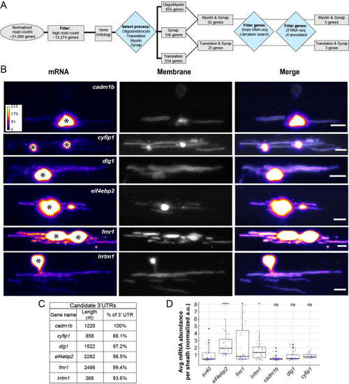
(A) Work flow to identify 3′ UTR candidates from RNA-seq data [27,43,44]. (B) Representative images from MS2 system showing localization of mRNAs containing different 3′ UTR sequences in oligodendrocytes. Asterisks mark cell bodies. Scale bars, 10 μm. (C) Table listing candidate 3′ UTRs incorporated into the MS2 system, 3′ UTR length, and the percentage of sequence that was cloned based on the annotated genome (GRCz11). (D) Average mRNA abundance, measured by average EGFP fluorescent intensity, per myelin sheath for each 3′ UTR. Normalized to sv40 control, statistical significance evaluated using Wilcoxon test. A minimum (n) of 5 larvae and 18 sheaths were used in each condition at 4 dpf. The underlying numerical data can be found in S1 and S2 Data. 3' UTR, 3' untranslated region; dpf, days post fertilization; RNA-seq, RNA sequencing.

<italic>eif4ebp2</italic> and <italic>fmr1</italic> mRNA are localized to sheaths during developmental myelination.

Representative images of smFISH experiments to visualize egfp, eif4ebp2, or fmr1 mRNA localization at 4 dpf (A and B) and 5 dpf (C and D) in sagittal sections of hindbrain (A, C) or transverse sections of the Mauthner axon in the spinal cord (B, D). Dashed lines outline cell bodies marked by EGFP-CAAX. Scale bars, 5 μm (A, C) or 1 μm (B, D). dpf, days post fertilization; smFISH, single molecule fluorescent in situ hybridization.

Common motifs in candidate 3′ UTRs are required for myelin localization.

(A) Schematic representation of the 3 motifs identified in the 3′ UTRs of mbpa-201, eif4ebp2-201, and fmr1-201 from the annotated zebrafish genome (GRCz11) using MEME suite bioinformatics software. (B) Schematic representation showing the relative position of the motifs within the 3′ UTRs. Green box is motif 1, blue box is motif 2, and red box is motif 3. Gray box is the conserved RTS previously identified as a minimal localization element necessary for Mbp mRNA transport in cultured oligodendrocytes [28,29,51]. Motif 1 in the mbpa 3′ UTR is not present in 3′ UTR isolated from zebrafish cDNA utilized in experimental procedures (3′ end of the dashed line). (C) Representative images of MS2 system after sequential deletions of all motifs from mbpa, eif4ebp2, and fmr1 3′ UTRs. Asterisks mark cell bodies. (D) Quantification of mRNA abundance in myelin sheaths from sequential deletions in (C). (E) Quantification of mRNA abundance in myelin sheaths from full length 3′ UTR, mbpa 3′ UTR variant 3–1177, or individual motif deletions. Statistical analysis evaluated with Wilcoxon test. Scale bars, 10 μm. A minimum (n) of 6 embryos and 35 sheaths were used in each condition (D and E). The underlying numerical data can be found in S1 and S4 Data. 3' UTR, 3' untranslated region; MEME, Multiple Em for Motif Elicitation; RTS, RNA transport signal.

Motif 2 is enriched in the mouse myelin transcriptome.

(A) Schematic representation of motifs 1, 2, or 3 enrichment in the myelin transcriptome in comparison to length-matched, randomized sequences using MEME suite Analysis of Motif Enrichment (version 5.1.1) [59]. (B) FIMO (version 5.1.1) was used to determine the frequency at which motif 2 is present in the myelin transcriptome [60]. cDNA sequences from the myelin transcriptome were analyzed for the presence of motif 2. One or more copies of motif 2 were present in 42.4% of myelin cDNAs. (C) FIMO was used to determine the frequency at which motif 2 is present in the mouse transcriptome. cDNA sequences from the mouse annotated genome (mm10) were analyzed for the presence of motif 2. One or more copies of motif 2 were present in 28.7% of mouse cDNAs. (D) Percentage of motif 2 occurrences in 5′ UTR, coding sequences, 3′ UTR or other positions in myelin transcriptome. Motifs in the “other” category represent motifs overlapping 2 regions. (E) Top 20 GO terms identified in the myelin transcriptome. (F) Top 20 GO terms identified in myelin transcripts containing motif 2. Terms are ordered from most to least significant based on -log2 of the false discovery rates. Counts represent number of genes identified with the GO term. The underlying numerical data can be found in S8S10 Data. FIMO, Find Individual Motif Occurrences; GO, gene ontology; MEME, Multiple Em for Motif Elicitation; UTR, untranslated region.

Acknowledgments
This image is the copyrighted work of the attributed author or publisher, and ZFIN has permission only to display this image to its users. Additional permissions should be obtained from the applicable author or publisher of the image. Full text @ PLoS Biol.