FIGURE SUMMARY
Title

Mutant dlx3b disturbs normal tooth mineralization and bone formation in zebrafish

Authors
Pang, L., Zhang, Z., Shen, Y., Cheng, Z., Gao, X., Zhang, B., Wang, X., Tian, H.
Source
Full text @ Peer J.

Generation of the mutant <italic>dlx3b</italic> zebrafish line.

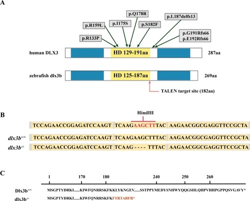
(A) Structure of the DLX3 and dlx3b proteins in human and zebrafish. The homeodomain and transactivation domains are marked in yellow and blue, respectively. The green arrows indicate the TDO mutation sites that found in patients. The red arrow indicates the TALEN target site in zebrafish. (B) Sequence of the TALEN target site and mutant site in zebrafish dlx3b. TALEN binding sites in the dlx3b gene are indicated in gray shading, and the HindIII restriction site is highlighted in red. The sequence of mutant dlx3b mRNA is aligned with wild-type dlx3b mRNA. (C) Predicted peptide sequences for the wild-type and mutant dlx3b. There is a frameshift mutation at 182 aa, and the subsequent mutant sequence is highlighted in red.

Identification and breeding of the mutant <italic>dlx3b</italic> zebrafish.

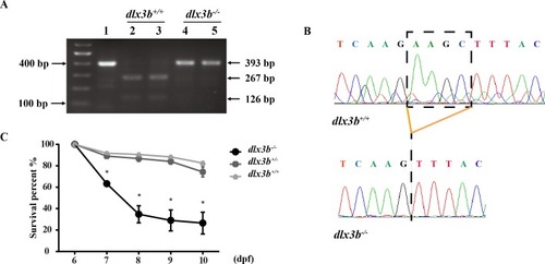
(A) Genotyping results identifying homozygote (dlx3b−∕−) mutant and wild-type siblings (dlx3b+∕+). Genomic DNA was amplified by PCR from the tails of the first-generation (F1) fish and then digested with a diagnostic restriction enzyme (Hind III). The first lane contains the original PCR product (393 bp). Lanes 2 and 3 contain wild-type siblings (dlx3b+∕+), and lanes 4 and 5 contain homozygotes (dlx3b−∕−). (B) Sequencing peak map of the DNA sample from the wild-type siblings (dlx3b+∕+) and mutant (dlx3b−∕−) zebrafish, respectively. The absence of the four-bp sequence (AAGC) in the mutant zebrafish (dlx3b−∕−) is clearly marked. (C) Homozygous mutant (dlx3b−∕−) zebrafish exhibited a lower survival rate compared with that of their wild-type (dlx3b+∕+) and heterozygous siblings (dlx3b+∕−) from 6–10 dpf. Within 5 d (6–9 dpf), the survival rate of the dlx3b−∕− group decreased from 100% to approximately 35%, while those of the dlx3b+∕+ group and dlx3b+∕− group decreased to 75%–80%.P < 0.05 vs. The dlx3b+∕+ group. n = 100 for each group (the starting population).

Body curvature of <italic>dlx3b</italic> homozygous mutant zebrafish.

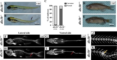
(A–B) Body curvature of 6–7 dpf larvae under a stereomicroscope. The body curvature in dlx3b−∕− (B) initially occurred in the early juvenile stages and was mainly limited to the distal end of the tail (black arrow), while the wild type (A) was showed no curvature. Scale bar: 300 µm. (C) Relative frequencies of dorsal curvature phenotypes in dlx3b+∕+ and dlx3b−∕− embryos. n = 100 for each group. (D–E) Mutant adult zebrafish (D) exhibited a severe curvature of the spine compared with wild-type siblings (E), especially in the tail part (black arrow). (F–I) Micro-CT results showing the spine from the lateral side (F & H) and ventral side (G & I). The abdominal and caudal spine of the dlx3b−∕− zebrafish developed a marked curvature in both the dorsal–ventral (G&I) and medial–lateral planes (F&H, red arrows). (J–K) Magnified imaging showing vertebral hypoplasia in the dlx3b−∕− zebrafish. A radiopaque area was observed in the caudal spine region (K, yellow arrow).

PHENOTYPE:
Fish:
Observed In:
Stage Range: Day 6 to Adult

Pharyngeal teeth of homozygous mutants (<italic>dlx3b</italic><sup>−∕−</sup>) exhibited deformiy.

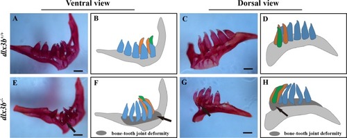
Compared with the pharyngeal teeth of the wild-type (A, C), those of the mutant group exhibited abnormal bending towards the dorsal side (E, G) with the abnormal bone growth of the pharyngeal bone at the bone-tooth joint (E, G, black arrow). (A–D) The pharyngeal dentition of the wild-type sibling; (E–H) the pharyngeal dentition of the dlx3b−∕− mutant. (A, E) The pharyngeal dentition viewed from the ventral side. (C, G) The pharyngeal dentition viewed from the dorsal side. (B, D, F, H) Schematic representations of A, C, E, and G, respectively (ventral tooth row, blue; mediodorsal tooth row, ochre; dorsal tooth row, green; replacement teeth not shown; and the abnormal bone growth is depicted as a dark gray region in F and H). Scale bar: 200 µm. n = 8 for each group.

Surfaces of the second and fourth ventral pharyngeal teeth of the <italic>dlx3b</italic> homozygous mutation zebrafish exhibited abnormal pits.

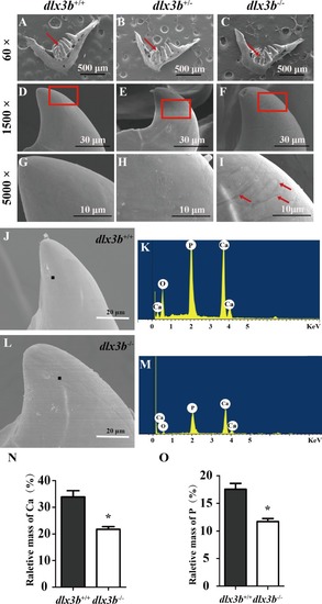
(A–I) SEM images showing the surface of the second pharyngeal tooth. The red arrows showed the teeth we studies below in a 60× image (A–C). The fourth ventral pharyngeal teeth (red arrows in A–C) was magnified as images D–F. The red rectangle areas in D–F were then magnified as G–I. The red arrows (I) indicate the pits on the surface of the second ventral pharyngeal tooth of the dlx3b−∕− fish. n=10 for each group (J–M) Lower relative mass fractions of mineral elements in the dlx3b−∕− mutant. The scanning areas for energy spectrum analysis were selected on the cusp of the pharyngeal teeth showed as images J and L. Energy spectrum of zebrafish pharyngeal teeth was showed as images K and M, mainly including O, Ca, and P. (N–O) The relative mass fraction of Ca (N) and P (O) significantly decreased in the dlx3b−∕− zebrafish compared with those in their wild-type siblings. n = 5, P < 0.001.

Down regulation of <italic>odam, scpp9, spp1</italic> and <italic>scpp5</italic> in <italic>dlx3b</italic><sup>−∕−</sup> fish.

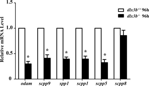
Real-time RT-PCR was used to determine the expression levels of mineralized-related genes using RNA samples extracted from zebrafish larvae at 96 hpf. The expression level of dlx3b+∕+ was set at 1 as a control, and the fold changes in dlx3b−∕− were calculated relative to dlx3b+∕+. P < 0.05 vs. the control. dlx3b+∕+, wild type siblings. dlx3b−∕−, mutant larvae.

EXPRESSION / LABELING:
Genes:
Fish:
Anatomical Term:
Stage: Day 4
PHENOTYPE:
Fish:
Observed In:
Stage: Day 4

Unillustrated author statements

PHENOTYPE:
Fish:
Observed In:
Stage Range: Day 6 to Days 7-13
Acknowledgments
This image is the copyrighted work of the attributed author or publisher, and ZFIN has permission only to display this image to its users. Additional permissions should be obtained from the applicable author or publisher of the image. Full text @ Peer J.