|
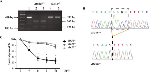
Identification and breeding of the mutant <italic>dlx3b</italic> zebrafish.(A) Genotyping results identifying homozygote (dlx3b−∕−) mutant and wild-type siblings (dlx3b+∕+). Genomic DNA was amplified by PCR from the tails of the first-generation (F1) fish and then digested with a diagnostic restriction enzyme (Hind III). The first lane contains the original PCR product (393 bp). Lanes 2 and 3 contain wild-type siblings (dlx3b+∕+), and lanes 4 and 5 contain homozygotes (dlx3b−∕−). (B) Sequencing peak map of the DNA sample from the wild-type siblings (dlx3b+∕+) and mutant (dlx3b−∕−) zebrafish, respectively. The absence of the four-bp sequence (AAGC) in the mutant zebrafish (dlx3b−∕−) is clearly marked. (C) Homozygous mutant (dlx3b−∕−) zebrafish exhibited a lower survival rate compared with that of their wild-type (dlx3b+∕+) and heterozygous siblings (dlx3b+∕−) from 6–10 dpf. Within 5 d (6–9 dpf), the survival rate of the dlx3b−∕− group decreased from 100% to approximately 35%, while those of the dlx3b+∕+ group and dlx3b+∕− group decreased to 75%–80%.∗P < 0.05 vs. The dlx3b+∕+ group. n = 100 for each group (the starting population).
|