FIGURE SUMMARY
Title

Polymeric nanobiotics as a novel treatment for mycobacterial infections

Authors
Batalha, I.L., Bernut, A., Schiebler, M., Ouberai, M.M., Passemar, C., Klapholz, C., Kinna, S., Michel, S., Sader, K., Castro-Hartmann, P., Renshaw, S.A., Welland, M.E., Andres Floto, R.
Source
Full text @ J. Control Release

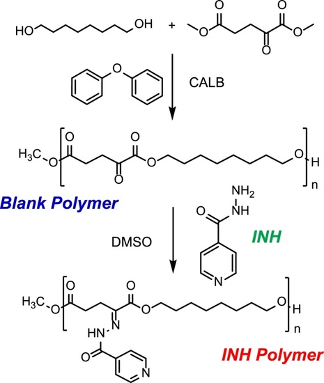
Synthesis of an α-keto polyester by (trans)esterification reaction catalysed by CALB and conjugation to isoniazid (INH).

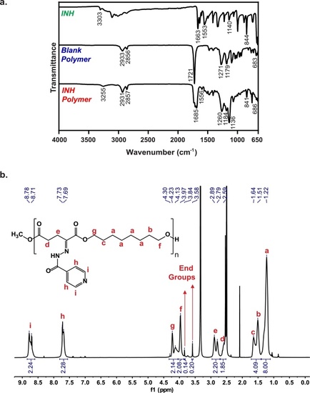
Characterization of polymer-drug conjugates. a. FTIR spectra of INH (top), Blank Polymer (middle), and INH Polymer (Bottom). b. 1H-NMR spectrum of INH Polymer in DMSO-d6 and peak assignments.

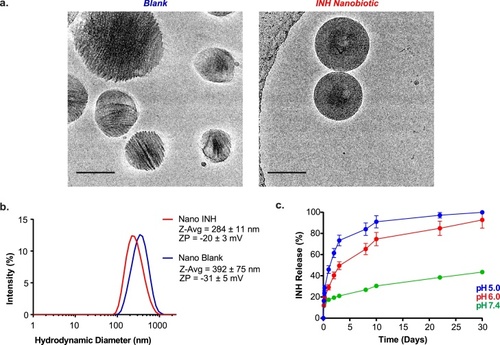
Characterization of polymeric nanobiotics a. Cryo-EM images show different shape and crystallinity of the Blank (left) and INH (right) nanobiotics (scale bar, 100 nm). b. Size distribution (nm) and zeta potential (mV) of Blank (blue) and INH (red) polymeric nanobiotics formulated by single emulsion solvent evaporation technique (n = 3). c. INH release from Nano INH at pH 5 (blue), 6 (red), and 7.4 (green), mimicking the acidic conditions of the phagolysosome and physiological conditions of systemic circulation (n = 3). (For interpretation of the references to colour in this figure legend, the reader is referred to the web version of this article).

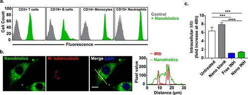
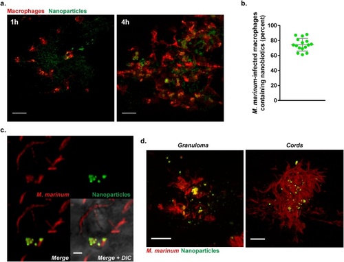
Nanobiotic uptake by phagocytic cells and in vitro efficacy against M. tuberculosis.a. Nanobiotics uptake by white blood cells quantified by fluorescence-activated cell sorting (FACS). Coumarin 6-labelled Nano INH are preferentially uptaken by phagocytic cells, namely monocytes (CD14+) and neutrophils (CD15+), rather than lymphocytes, such as T cells (CD3+) and B cells (CD19+), likely reflecting their large size distribution. b. Confocal microscopy images of differentiated THP-1 cells infected with a mCherry fluorescent reporter strain of M. tuberculosis H37Rv ΔleuD ΔpanCD (red) and treated with Cou-6-labelled Nano INH (green) (scale bar, 10 μm) c. Differentiated THP-1 cells were infected with a luminescent reporter strain M. tuberculosis H37Rv ΔleuD ΔpanCD, treated with 100 μM INH either as a free drug or as nanodrug and intracellular M. tuberculosis was assessed 48 h post-infection by relative luminescence units (RLUs). Untreated cells and cells treated with drug-free nanobiotics (Nano Blank) were used as negative controls. Results are presented in terms of RLUs normalized to untreated cells (Mean ± SEM, n = 6). (For interpretation of the references to colour in this figure legend, the reader is referred to the web version of this article).

In vivo testing of multi-drug nanobiotics in a M. marinum-infected zebrafish larval model. a. Confocal microscopy images showing nanobiotics-induced macrophage mobilization in vivo. Suspension of coumarin 6-labelled Nano Blank (green) was injected into the muscle of 3 dpf Tg(mpeg1:mCherryCAAX)sh378 zebrafish line harbouring red macrophages. Macrophage chemotaxis towards injection site has been monitored at 1 and 4 h post injection (scale bar, 20 μm). b. Quantification and c. Confocal imaging of coumarin 6-labelled Nano Blank (green) uptake by M. marinum-infected macrophages (red) after 4 h post infection (scale bar, 1 μm). d. Confocal imaging showing the repartition and accumulation of coumarin 6-labelled Nano Blank (green) into a M. marinum (red)-granuloma (left, scale bar, 5 μm) and a mycobacterial cord structure (right, scale bar, 5 μm). (For interpretation of the references to colour in this figure legend, the reader is referred to the web version of this article).

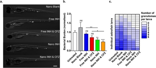
Effect of nanobiotics at 3 days post infection on zebrafish infected with fluorescently-labelled M. marinum.a. representative images (scale bar, 200 μm). b. quantification of bacterial load (results plotted as mean ± SEM from 2 independent experiments; n = 21). c. Quantification of granuloma number at 3dpi. Results are plotted as mean ± SEM from 2 independent experiments (n = 19).

Acknowledgments
This image is the copyrighted work of the attributed author or publisher, and ZFIN has permission only to display this image to its users. Additional permissions should be obtained from the applicable author or publisher of the image. Full text @ J. Control Release