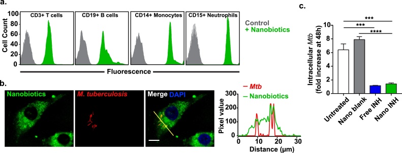
IMAGE

Fig. 4

ID
ZDB-IMAGE-200124-57
Source
Figures for Batalha et al., 2019
Image
Figure Caption

Fig. 4

Nanobiotic uptake by phagocytic cells and in vitro efficacy against M. tuberculosis.a. Nanobiotics uptake by white blood cells quantified by fluorescence-activated cell sorting (FACS). Coumarin 6-labelled Nano INH are preferentially uptaken by phagocytic cells, namely monocytes (CD14+) and neutrophils (CD15+), rather than lymphocytes, such as T cells (CD3+) and B cells (CD19+), likely reflecting their large size distribution. b. Confocal microscopy images of differentiated THP-1 cells infected with a mCherry fluorescent reporter strain of M. tuberculosis H37Rv ΔleuD ΔpanCD (red) and treated with Cou-6-labelled Nano INH (green) (scale bar, 10 μm) c. Differentiated THP-1 cells were infected with a luminescent reporter strain M. tuberculosis H37Rv ΔleuD ΔpanCD, treated with 100 μM INH either as a free drug or as nanodrug and intracellular M. tuberculosis was assessed 48 h post-infection by relative luminescence units (RLUs). Untreated cells and cells treated with drug-free nanobiotics (Nano Blank) were used as negative controls. Results are presented in terms of RLUs normalized to untreated cells (Mean ± SEM, n = 6). (For interpretation of the references to colour in this figure legend, the reader is referred to the web version of this article).

Acknowledgments
This image is the copyrighted work of the attributed author or publisher, and ZFIN has permission only to display this image to its users. Additional permissions should be obtained from the applicable author or publisher of the image. Full text @ J. Control Release