
- Title
-
αKlotho Regulates Age-Associated Vascular Calcification and Lifespan in Zebrafish
- Authors
- Singh, A.P., Sosa, M.X., Fang, J., Shanmukhappa, S.K., Hubaud, A., Fawcett, C.H., Molind, G.J., Tsai, T., Capodieci, P., Wetzel, K., Sanchez, E., Wang, G., Coble, M., Tang, W., Cadena, S.M., Fishman, M.C., Glass, D.J.
- Source
- Full text @ Cell Rep.
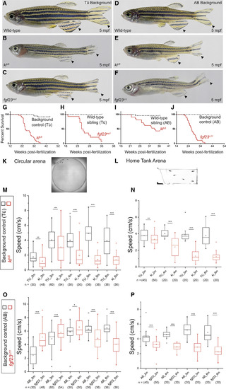
Zebrafish αklotho and fgf23 Mutants (A–F) Body condition of αklotho and fgf23 mutant males at 5 mpf: (A) Tü wild-type strain, (B) αklotho (klΔ5), and (C) fgf23 (fgf23ins1) mutant in Tü background; (D) AB wild-type strain, (E) αklotho (klΔ5), and (F) fgf23 (fgf23Δ11) mutant in AB background. (G–J) Survival curves for (G) αklotho (n = 36 background controls, 32 mutants; p < 0.0001) and (H) fgf23 mutants (n = 14 wild-type siblings, 14 mutants; p < 0.0001) in Tü background and for (I) αklotho (n = 24 wild-type siblings, 21 mutants; p = 0.0001) and (J) fgf23 mutants (n = 60 background controls, 68 mutants; p < 0.0001) in AB background. Log-rank (Mantel-Cox) test for statistical analysis on survival curves in GraphPad Prism. (K and L) Analysis of speed (cm) in the (K) circular arena and (L) home-tank arena. Age (m; mpf) on x axis; n = number of fish. (M and N) αklotho mutants and wild-type controls in (M) circular and (N) home-tank arena. (O and P) fgf23 mutants and wild-type controls in (O) circular and (P) home-tank arena. Statistical analysis using unpaired t test in GraphPad Prism. PHENOTYPE:
|
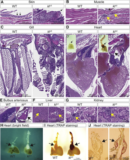
Vascular Calcification and Inflammation in αklothoMutants H&E staining on paraffin sections from 5-month-old wild-type control (Tü) and αklotho (klΔ5) males. Shown are (A) skin (arrows indicate mucous cells in wild type); (B) muscle (arrows indicate vascular calcification); (C) gills; (D) heart (arrow indicates calcification in the BA); (D′ and D″) alizarin red-stained whole-mount hearts (arrows indicate the BA); (E) BA (arrow indicates calcification); (F) liver (arrows indicate bile-duct); and (G) kidney (arrows indicate glomeruli). (H) Bright-field images of 5-mpf wild-type (left) and klΔ5 (right) hearts. TRAP staining on (I) whole mount and (J) cryosection of 5-mpf hearts. PHENOTYPE:
|

ZFIN is incorporating published figure images and captions as part of an ongoing project. Figures from some publications have not yet been curated, or are not available for display because of copyright restrictions. |
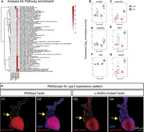
Pathway Enrichment Analysis of Differentially Upregulated Genes (A) Gene set enrichment analysis comparing upregulated pathways by αklotho mutation in the kidney, heart, and gills. Each row is a pathway, annotated on the right-hand side, and each column corresponds to the significance of the enrichment analysis for each tissue and adult stage (m3, 3 mpf; m5, 5 mpf). The colors from white to red represent the negative log10 adjusted p value from low to high. Only pathways that were enriched significantly (adjusted p value < 0.05) in at least one tissue were included. (B–G) Box plots showing select examples of genes upregulated at 5 mpf in αklotho mutant hearts (blue, kl) compared to wild-type (WT) siblings (red). Shown are (B) mmp9 (matrix metallopeptidase 9), (C) mmp13a (matrix metallopeptidase 13a), (D) entpd5a (ectonucleoside triphosphate diphosphohydrolase 5a), (E) spp1 (secreted phosphoprotein 1), (F) ctsk (cathepsin K), and (G) acp5a (acid phosphatase 5a, tartrate resistant). Age: 3 and 5 mpf. (H) RNAscope for spp1: (H1 and H2) WT control; (H3 and H4) αklotho (klΔ5) mutant hearts stained with DAPI (blue), and spp1RNAscope probe (red). White lines outline the BA (arrow) and the blood vessel leading to gills. |
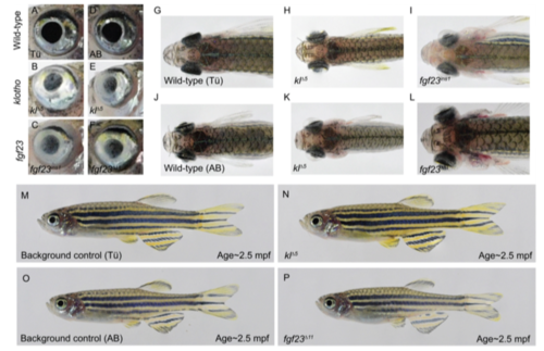
Phenotypic characterization of zebrafish αklotho and fgf23mutants. (A-F) close-up of an eye with opaque overgrowth in αklotho, and fgf23 mutant females: (A) Tü wildtype strain, (B) αklotho, and (C) fgf23 mutant in Tü background; (D) AB wildtype strain, (E) αklotho, and (F) fgf23mutant in AB background. (G-L) top-view showing eye protrusion in αklotho and fgf23 mutant females: (G) Tü wildtype strain, (H) αklotho, and (I) fgf23 mutant in Tü background; (J) AB wildtype strain, (K) αklotho, and (L)fgf23 mutant in AB background. (M-P) αklotho and fgf23 mutants appear comparable to background controls at 2.5 mpf: (M) wildtype and, (N) αklotho (klΔ5) males in Tü background; (O) wildtype and (P) fgf23 (fgf23Δ11) males in AB background. |

Histological characterization of zebrafish heart. (A) H&E-stained serial sections across caudal bone of wildtype and αklotho mutant; dashed yellow lines demarcate region enlarged in inset. Genotypes as indicated in the image (n=3 males, 5 months post-fertilization). (B) Alizarin red staining on fgf23 mutant hearts to visualize calcification. Alizarin red staining on whole-mount hearts of background control (AB; left) and fgf23 mutant (fgf23Δ11; right). Intense red staining is observed in the outflow tract of the heart (arrow) in fgf23 mutant. (n=5 males, 5 months post-fertilization). |