
- Title
-
Genetic disruption of 21-hydroxylase in zebrafish causes interrenal hyperplasia
- Authors
- Eachus, H., Zaucker, A., Oakes, J.A., Griffin, A., Weger, M., Güran, T., Taylor, A., Harris, A., Greenfield, A., Quanson, J.L., Storbeck, K.H., Cunliffe, V.T., Müller, F., Krone, N.
- Source
- Full text @ Endocrinology
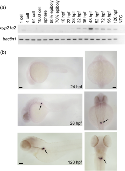
Zebrafish cyp21a2 expression is largely restricted to the interrenal gland. (a) Analysis of cyp21a2 expression during embryonic and early larval development by RT-PCR with b-actin1 as an internal standard (gel images inversed). A no-template control sample (NTC) served as negative control. The onset of detectable cyp21a2 expression is at 28 hpf, after which cyp21a2 continues to be expressed at all stages examined. (b) Images of wild-type larvae at different stages after WISH against cyp21a2. Left panels show lateral view, with head to the left; right panels show a dorsal view. No staining is seen in 24-hpf embryos. In 28-hpf embryos and 120-hpf larvae, strong staining is observed in the interrenal gland region, with some faint signal detected at 120 hpf in the head (arrows). Scale bar: 0.1 mm. |
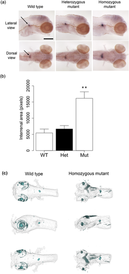
Zebrafish cyp21a2 mutants have enlarged interrenal tissue at 120 hpf. (a) Expression of cyp17a2 in 120-hpf cyp21a2uob2122 wild-type, heterozygous, and homozygous cyp21a2uob2122 mutant larvae in lateral (upper panel) and dorsal (lower panel) views. The area of cyp17a2-positive interrenal tissue (arrows) is enlarged in homozygous mutants. n = 6 each. Scale bar: 0.25 mm. (b) Quantification of the area of cyp17a2-positive interrenal tissue in 120-hpf cyp21a2uob2122 larvae. The interrenal tissue is significantly larger in homozygous mutants (Mut) than in wild-type (WT) and heterozygous (Het) siblings (ANOVA: F = 12.15, df = 2,15, P = 0.0007; Tukey: WT vs. Het P = 0.895, WT vs. Mut P = 0.003, Het vs. Mut P = 0.003). **P < 0.01 compared with wild types and heterozygotes. n = 4 to 8 each. (c) OPT imaging of 120-hpf cyp21a2 larvae after WISH for cyp17a2 reveals an enlarged interrenal gland in cyp21a2uob2122 homozygous mutants (right) compared with wild-type siblings (left). Whole-mount ventral (upper), lateral (middle), and dorsal (lower) views are shown. |

ZFIN is incorporating published figure images and captions as part of an ongoing project. Figures from some publications have not yet been curated, or are not available for display because of copyright restrictions. PHENOTYPE:
|
|
ZFIN is incorporating published figure images and captions as part of an ongoing project. Figures from some publications have not yet been curated, or are not available for display because of copyright restrictions. |
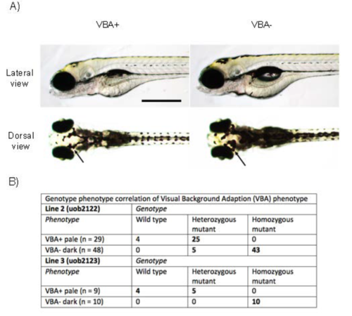
Zebrafish cyp21a2 mutants have an impaired Visual Background Adaption (VBA) phenotype A) Lateral (upper panel) and dorsal (lower panel) view images of 120 hpf larvae from a cyp21a2uob2122/+ in-cross. Overall morphology is comparable, but, unlike VBA+ larvae, VBA- larvae fail to aggregate the melanosomes in their melanophores when exposed to bright light; compare melanophores (arrows) between VBA+ and VBA- larva. Scale bar: 0.5 mm. B) Genotyping of cyp21a2uob2122 and cyp21a2uob2123 larvae after VBA assay. The VBA+ larvae are always wild types or heterozygotes. The VBA- larvae are almost always homozygous mutants. PHENOTYPE:
|
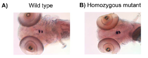
Zebrafish cyp21a2 mutants have an enlarged pituitary gland In situ hybridisation for pomca reveals a larger field of expression in the anterior and posterior pituitary of cyp21a2uob2122 homozygous mutant larvae at 120 hpf. Ventral whole-mount view. |