
- Title
-
Construction and characterization of a sox9b transgenic reporter line.
- Authors
- Plavicki, J.S., Baker, T.R., Burns, F.R., Xiong, K.M., Gooding, A.J., Hofsteen, P., Peterson, R.E., Heideman, W.
- Source
- Full text @ Int. J. Dev. Biol.
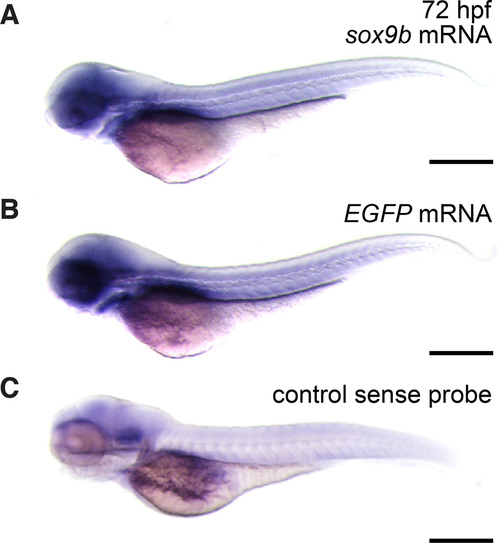
sox9b transgenic line recapitulates the endogenous pattern of em>sox9b expression (A–C) Lateral views of 72 hpf embryos. To detect sox9b, in situ hybridization was performed on whole embryos. (A) sox9b mRNA in transgenic embryos. (B) EGFP mRNA in transgenic embryos. (C) A sense probe was used as a control for nonspecific binding. Scale bars, 100 microns. EXPRESSION / LABELING:
|
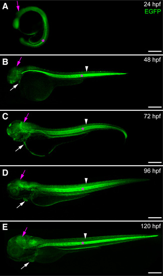
Expression of sox9b:EGFP during embryonic and larval development (A–E) Lateral views of sox9b:EGFP embryos and larvae. (A) Epifluorescent image at 24 hpf. (B–E) Confocal images at 48 hpf (B), 72 hpf (C), 96 hpf (D) and 120 hpf (E). sox9b:EGFP expression is detected in the brain (purple arrows), eye, heart (white arrows), jaw, spinal cord (white arrowhead) and notocord (pink asterisks). Scale bars, 100 microns. |
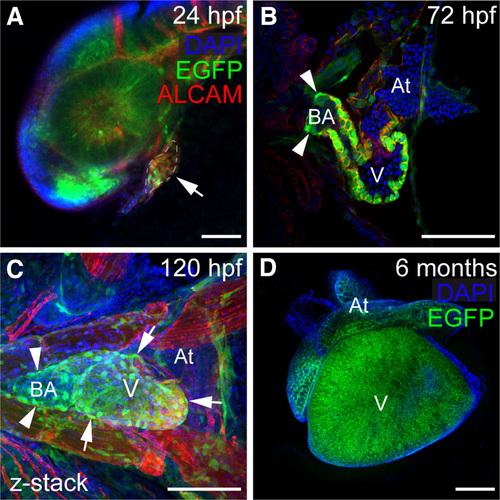
Expression of sox9b:EGFP in the heart (A–D) Confocal images of sox9b:EGFP expression in embryonic, larval, and adult zebrafish hearts. Cardiomyocytes are marked with activated leukocyte cell adhesion molecule (ALCAM; red in A–C) and nuclei are stained with DAPI (blue); (A) Lateral view of a 24 hpf embryo. The developing heart is outlined and indicated by the arrow. (B,C) Ventral views of a 72 and 96 hpf hearts. Expression of sox9b:EGFP is detected in cardiomyocytes, the bulbus arteriosus (arrowheads in B and C), and in epicardial cells (arrows in C). (D) Ventral view of an adult heart. Expression of sox9b:EGFP in cardiomyocytes continues into adulthood. V is ventricle; At, atrium; BA, bulbus arteriosus. Anterior is to the left in all panels. Scale bars, 50 microns. |
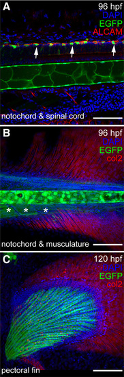
Expression of sox9b:EGFP in larval spine, skeleton, and fin (A–C) Lateral views of a 96 hpf and 120 larvae. Confocal images of sox9b:EGFP expression (green) in zebrafish spinal cord (A), notochord (A,B), and pectoral fin (C). (A) Neurons in the spinal cord are marked with activated leukocyte cell adhesion molecule (ALCAM; red in A). Expression of sox9b:EGFP is seen in a subset of neurons (arrows) and in the notochord. (B) sox9b:EGFP is also expressed in muscle associated with the spine (asterisks in B) and the developing pectoral fin. Collagen 2 (col2; red in B,C) is expressed in the fins. (A–C) Nuclei are stained with DAPI (blue). Scale bars, 50 microns. |
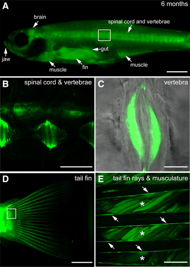
Expression of sox9b:EGFP in adult skeleton and skeletal muscle (A–E) Lateral views of six month old sox9b:EGFP transgenic fish. (B) Area boxed in (A). Expression is detected in the spinal cord and vertebrae. (C) Magnification of the sox9b:EGFP expressing growth plates in the vertebrae. (D) Muscle associated with the tail fin and rays within the tail fin express sox9b:EGFP. (E) Magnification of the box in (D). Asterisks mark the muscle while arrows point to the rays. Scale bars in (A,D), 150 microns. Scale bars in (B,C,E), 50 microns. EXPRESSION / LABELING:
|
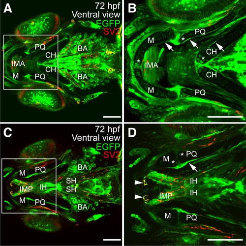
Expression of sox9b:EGFP in larval jaw and associated skeletal muscle (A–D) Ventral views of 72 hpf larvae stained for synaptic vesicles (SV2; red). sox9b:EGFP expression was detected in the perichondrium and in chondrocytes. (B,D) Magnification of regions in the white boxes in panels (A,C) respectively. Expression is associated with the ceratohyal (CH), Meckel’s (M), palatoquadrate (PQ), ceratobranchials (not shown) and basihyal (not shown) cartilages. Expression was also detected in muscles associated with the jaw including the intermandibularis anterior (IMA), intermandibularis posterior (IMP), interhyoideus (IH), and sternohyoideus (SH). sox9b:EGFP was also observed in the branchial arches (BA). Scale bars, 50 microns. EXPRESSION / LABELING:
|
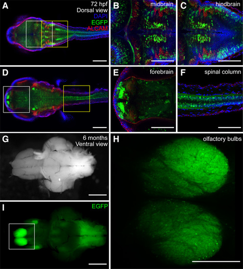
Expression of sox9b:EGFP in larval and adult brain (A–F) Confocal images showing dorsal views of a 72 hpf larval zebrafish. Larvae were stained for ALCAM (red) and mounted in Vectashield with DAPI (blue). (B,C) Magnification of the regions in the white and yellow boxes in (A). (E,F) Magnification of the regions in the white and yellow boxes in (D). Expression of sox9b:EGFP in the fore-, mid- and hindbrain as well as in the spinal cord. (G,I) Bright field images and epifluorescence and ventral views of an adult brain. (H) Magnification of olfactory bulb boxed in (I). Scale bars, 50 microns. EXPRESSION / LABELING:
|
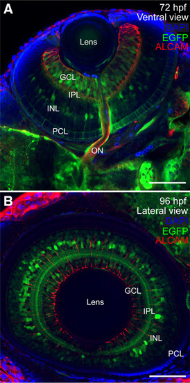
Expression of sox9b:EGFP in retina (A,B) Ventral and lateral views of larval eyes at 72 and 96 hpf stained for ALCAM (red) and mounted in Vectashield with DAPI (blue). (A) At 72 hpf, sox9b:EGFP expression was observed in a subset of cells in the photoreceptor cell layer (PCL), inner nuclear layer (INL), and ganglion cell layer (GCL) and within the inner plexiform layer (IPL). (B) By 96 hpf, sox9b:EGFP expression in the PCL was waning, however sox9b:EGFP was visible in the INL, IPL, and GCL. Scale bars, 50 microns. EXPRESSION / LABELING:
|
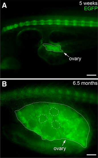
Expression of sox9b:EGFP in ovary Lateral views of juvenile (A) and adult (B) ovary. The ovaries are outlined with a solid line. Examples of mature follicles are outlined with dashed circles. Scale bars, 100 microns. EXPRESSION / LABELING:
|

ZFIN is incorporating published figure images and captions as part of an ongoing project. Figures from some publications have not yet been curated, or are not available for display because of copyright restrictions. |
|
Unillustrated author statements EXPRESSION / LABELING:
|