
Fig. 7
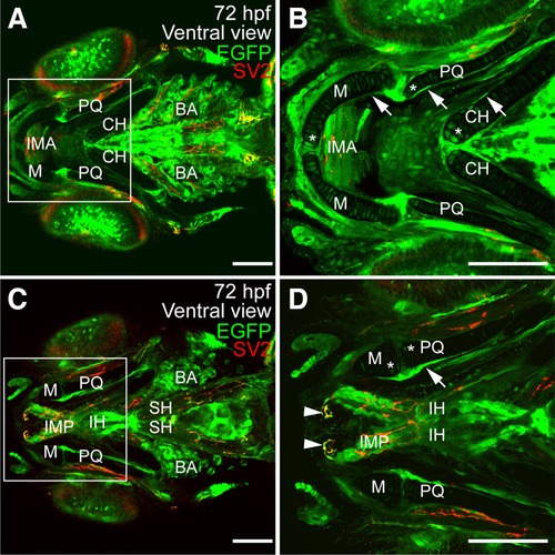
Expression of sox9b:EGFP in larval jaw and associated skeletal muscle (A–D) Ventral views of 72 hpf larvae stained for synaptic vesicles (SV2; red). sox9b:EGFP expression was detected in the perichondrium and in chondrocytes. (B,D) Magnification of regions in the white boxes in panels (A,C) respectively. Expression is associated with the ceratohyal (CH), Meckel’s (M), palatoquadrate (PQ), ceratobranchials (not shown) and basihyal (not shown) cartilages. Expression was also detected in muscles associated with the jaw including the intermandibularis anterior (IMA), intermandibularis posterior (IMP), interhyoideus (IH), and sternohyoideus (SH). sox9b:EGFP was also observed in the branchial arches (BA). Scale bars, 50 microns. |
| Gene: | |
|---|---|
| Fish: | |
| Anatomical Terms: | |
| Stage: | Protruding-mouth |