
Fig. 5
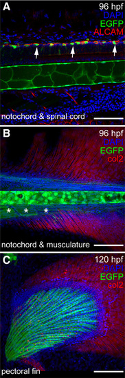
Expression of sox9b:EGFP in larval spine, skeleton, and fin (A–C) Lateral views of a 96 hpf and 120 larvae. Confocal images of sox9b:EGFP expression (green) in zebrafish spinal cord (A), notochord (A,B), and pectoral fin (C). (A) Neurons in the spinal cord are marked with activated leukocyte cell adhesion molecule (ALCAM; red in A). Expression of sox9b:EGFP is seen in a subset of neurons (arrows) and in the notochord. (B) sox9b:EGFP is also expressed in muscle associated with the spine (asterisks in B) and the developing pectoral fin. Collagen 2 (col2; red in B,C) is expressed in the fins. (A–C) Nuclei are stained with DAPI (blue). Scale bars, 50 microns. |
| Gene: | |
|---|---|
| Fish: | |
| Anatomical Terms: | |
| Stage Range: | Day 4 to Day 5 |