
Image
|
Figure Caption
Fig. 9
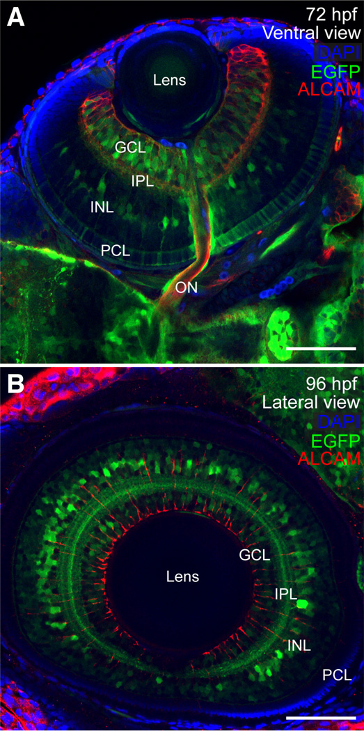
Expression of sox9b:EGFP in retina
(A,B) Ventral and lateral views of larval eyes at 72 and 96 hpf stained for ALCAM (red) and mounted in Vectashield with DAPI (blue). (A) At 72 hpf, sox9b:EGFP expression was observed in a subset of cells in the photoreceptor cell layer (PCL), inner nuclear layer (INL), and ganglion cell layer (GCL) and within the inner plexiform layer (IPL). (B) By 96 hpf, sox9b:EGFP expression in the PCL was waning, however sox9b:EGFP was visible in the INL, IPL, and GCL. Scale bars, 50 microns.
Figure Data
Acknowledgments
This image is the copyrighted work of the attributed author or publisher, and
ZFIN has permission only to display this image to its users.
Additional permissions should be obtained from the applicable author or publisher of the image.
Full text @ Int. J. Dev. Biol.