
- Title
-
TFAP2 paralogs regulate melanocyte differentiation in parallel with MITF
- Authors
- Seberg, H.E., Van Otterloo, E., Loftus, S.K., Liu, H., Bonde, G., Sompallae, R., Gildea, D.E., Santana, J.F., Manak, J.R., Pavan, W.J., Williams, T., Cornell, R.A.
- Source
- Full text @ PLoS Genet.
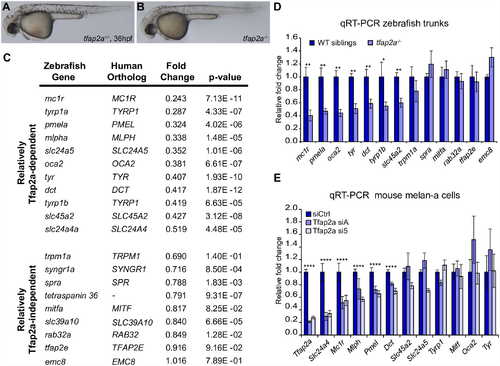
Melanocyte differentiation genes are sensitive to the loss of TFAP2A. (A, B) Lateral images of tfap2a+/- and tfap2a-/- mutant zebrafish at 36 hpf. tfap2a-/- mutants have fewer melanocytes and delayed pigmentation relative to their wildtype or tfap2a+/- siblings. (C) Fold changes calculated from microarray expression levels for 20 zebrafish melanocyte genes. Top: 11 genes with expression levels between 0.2–0.55-fold in tfap2a-/- mutants compared to WT siblings. Bottom: 9 genes with expression levels reduced no further than the number of melanocytes in tfap2a-/- mutants. (D) qRT-PCR validation of zebrafish microarray results for 13 melanocyte genes (Mann-Whitney U, *p<0.05, **p<0.01). (E) qRT-PCR validation of microarray results for 12 melanocyte genes in mouse melan-a cells transfected with control siRNA or each of two siRNAs targeting Tfap2a (siA, si5). Significance evaluated only when both siA and si5 altered gene expression in the same direction relative to siCtrl (one-way ANOVA with Bonferroni correction, ****p<0.0001). |
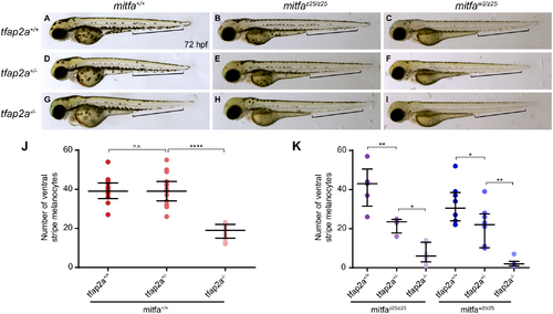
tfap2a and mitfa display a genetic interaction in zebrafish. (A-I) Compared to wildtype embryos at 72 hpf (A), melanocytes in mitfaz25/z25 mutant embryos (B) are dark but punctate, and melanocytes in mitfaw2/z25 trans-heterozygous embryos (C) are pale, punctate, and reduced in number. While heterozygous mutation of tfap2a in the wildtype background (tfap2a+/-) (D) does not result in a melanocyte phenotype, mitfaz25/z25;tfap2a+/- mutants (E) and mitfaw2/z25;tfap2a+/- mutants (F) display a loss of melanocytes in the ventral stripe (brackets). Similarly, the well-characterized phenotype of fewer, paler melanocytes in tfap2a-/- null mutants (G) is more pronounced in both the mitfaz25/z25 mutant background (H) and the mitfaw2/z25 mutant background (I). (J, K) Counts of ventral stripe melanocytes (see brackets in A-I) showed no difference between wildtype and tfap2a+/- embryos (J, n = 10+), while tfap2a+/- embryos in both mitfa mutant backgrounds had significantly fewer melanocytes in the ventral stripe (K, n = 4–8). One-way ANOVA: *p<0.05, **p<0.01, ****p<0.0001. PHENOTYPE:
|
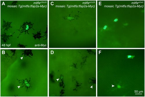
Forced expression of tfap2a does not rescue melanocytes in mitfa mutant zebrafish. (A-D) Nuclei of melanocytes expressing mosaic Tg(mitfa:tfap2a-Myc) are brightly labeled after anti-Myc immunostaining. At 48 hpf, labeled melanocytes in mitfaz25/z25 mutant embryos (A, B) and mitfaw2/z25 mutant embryos (C, D) display no apparent improvement in pigmentation or dendricity compared to adjacent unlabeled cells (B and D, white arrowheads). (E, F) In mitfaw2/w2 mutants, all labeled cells are unpigmented and resemble other cell types including xanthophores (F, white arrowhead). |
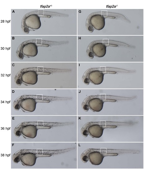
Time course of delayed melanocyte differentiation in tfap2a-/- zebrafish. (A-L) Lateral views of live tfap2a+/- (left column) and tfap2a-/- (right column) zebrafish embryos at the indicated age from 28-38 hpf. (C, I) Melanocytes in a tfap2a+/- zebrafish (C) appear darkly pigmented by 32 hpf, whereas melanocytes at the same location in a tfap2a-/- mutant (I) at this stage remain pale and punctate. (F, L) At 38 hpf, there is still a detectable difference in the level of pigmentation in tfap2a+/- (F) and tfap2a-/- (L) mutant animals. |