
- Title
-
Mycobacterial WhiB6 Differentially Regulates ESX-1 and the Dos Regulon to Modulate Granuloma Formation and Virulence in Zebrafish
- Authors
- Chen, Z., Hu, Y., Cumming, B.M., Lu, P., Feng, L., Deng, J., Steyn, A.J., Chen, S.
- Source
- Full text @ Cell Rep.
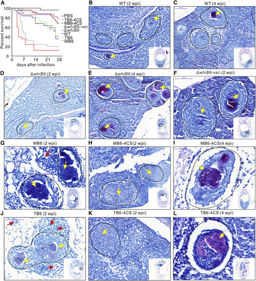
Holo- and Reduced Apo-WhiB6 Modulate Virulence of Mm and Granuloma Necrosis in Adult Zebrafish (A) Kaplan-Meier graph showing survival of adult zebrafish injected intraperitoneally with ~5,000 cfu the indicated Mm strains or an equivalent volume of PBS. n = 25 for each group. Survival was compared by log-rank test: MB6 versus ΔwhiB6-vec, MB6 versus MB6-4CS, and TB6 versus TB6-4CS, p < 0.0001; TB6 versus ΔwhiB6-vec, p < 0.001; TB6-4CS versus ΔwhiB6-vec, p < 0.05; MB6-4CS versus ΔwhiB6-vec,p = 0.0727; and WT versus ΔwhiB6, p = 0.6633. (B-E) Representative Ziehl-Neelsen stains of adult zebrafish infected with WT (B and C) or ΔwhiB6 (D and E) at 2 weeks (B and D) or 4 weeks (C and E) after infection, respectively. Scale bars, 100 µm. (F-H, J, and K) Representative Ziehl-Neelsen stains of adult zebrafish 2 weeks postinfection with Mm strains. Scale bars, 100 µm. (I and L) Representative Ziehl-Neelsen stains of adult zebrafish 4 weeks postinfection with MB6-4CS (I) and TB6-4CS (L). Scale bars, 50 µm. Dotted circles delineate granulomas. Yellow arrows indicate mycobacteria inside granulomas, and red arrows indicate mycobacteria outside granulomas. Three fish per group were used for each time point. PHENOTYPE:
|
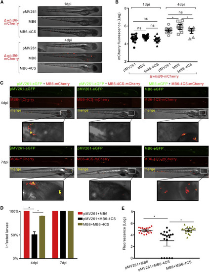
Holo-WhiB6 Accelerates Replication and Spread of Mm in Zebrafish Larvae (A and B) Representative images (A) and mean bacterial burden (B) of zebrafish larvae infected with ~150 cfu fluorescent Mm strains via the caudal vein at 1 and 4 dpi. Scale bar, 70 µm (A). Error bars indicate SEM (B). (C) Representative images of larvae co-infected with ~150 cfu each of two of the fluorescent Mm strains via the caudal vein at 4 and 7 dpi. Scale bar, 70 µm. (D) Percentage of larvae with Mm in the brain at 4 and 7 dpi. Data represent the average values from two independent experiments. (E) Mean fluorescence of larvae co-infected with ~150 cfu each of two of the fluorescent Mm strains via the caudal vein at 7 dpi. Error bars indicate SEM of two independent experiments performed in triplicate. |
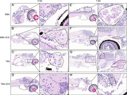
Histological observation of holo- and apo-reduced-like WhiB6 in modulating replication and spread of Mm cells in zebrafish larvae. Related to Figure 6. (A-H) Representative hematoxylin and eosin stains of zebrafish larvae 4 dpi (A-D) or 7 dpi (E-H) caudal infected with ~150 CFU of the indicated Mm strains. Areas enlarged are shown on the right of each image. Dotted circles delineate locations where Mm distributed, resulting in tissue damage and bacterial replication. Three fish per group were used for each time point. |