
|
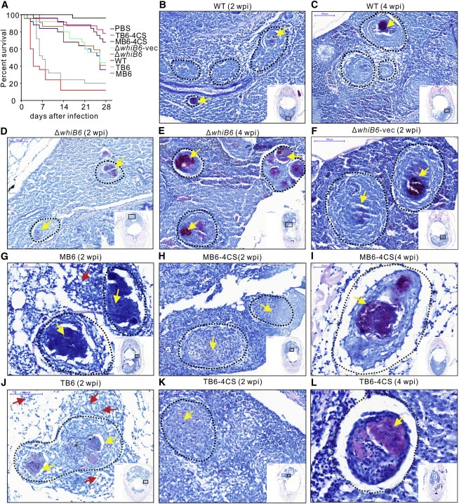
Fig. 5
Holo- and Reduced Apo-WhiB6 Modulate Virulence of Mm and Granuloma Necrosis in Adult Zebrafish
(A) Kaplan-Meier graph showing survival of adult zebrafish injected intraperitoneally with ~5,000 cfu the indicated Mm strains or an equivalent volume of PBS. n = 25 for each group. Survival was compared by log-rank test: MB6 versus ΔwhiB6-vec, MB6 versus MB6-4CS, and TB6 versus TB6-4CS, p < 0.0001; TB6 versus ΔwhiB6-vec, p < 0.001; TB6-4CS versus ΔwhiB6-vec, p < 0.05; MB6-4CS versus ΔwhiB6-vec,p = 0.0727; and WT versus ΔwhiB6, p = 0.6633.
(B-E) Representative Ziehl-Neelsen stains of adult zebrafish infected with WT (B and C) or ΔwhiB6 (D and E) at 2 weeks (B and D) or 4 weeks (C and E) after infection, respectively. Scale bars, 100 µm.
(F-H, J, and K) Representative Ziehl-Neelsen stains of adult zebrafish 2 weeks postinfection with Mm strains. Scale bars, 100 µm.
(I and L) Representative Ziehl-Neelsen stains of adult zebrafish 4 weeks postinfection with MB6-4CS (I) and TB6-4CS (L). Scale bars, 50 µm. Dotted circles delineate granulomas.
Yellow arrows indicate mycobacteria inside granulomas, and red arrows indicate mycobacteria outside granulomas. Three fish per group were used for each time point.