
|
Fig. 6
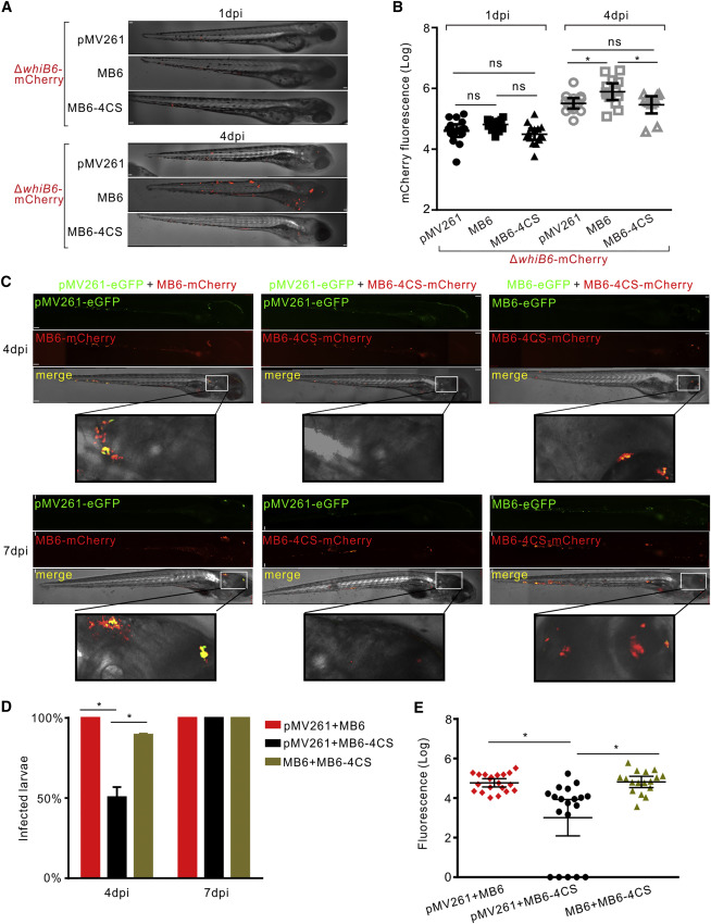
Holo-WhiB6 Accelerates Replication and Spread of Mm in Zebrafish Larvae
(A and B) Representative images (A) and mean bacterial burden (B) of zebrafish larvae infected with ~150 cfu fluorescent Mm strains via the caudal vein at 1 and 4 dpi. Scale bar, 70 µm (A). Error bars indicate SEM (B).
(C) Representative images of larvae co-infected with ~150 cfu each of two of the fluorescent Mm strains via the caudal vein at 4 and 7 dpi. Scale bar, 70 µm. (D) Percentage of larvae with Mm in the brain at 4 and 7 dpi. Data represent the average values from two independent experiments.
(E) Mean fluorescence of larvae co-infected with ~150 cfu each of two of the fluorescent Mm strains via the caudal vein at 7 dpi. Error bars indicate SEM of two independent experiments performed in triplicate.