
Image
|
Figure Caption
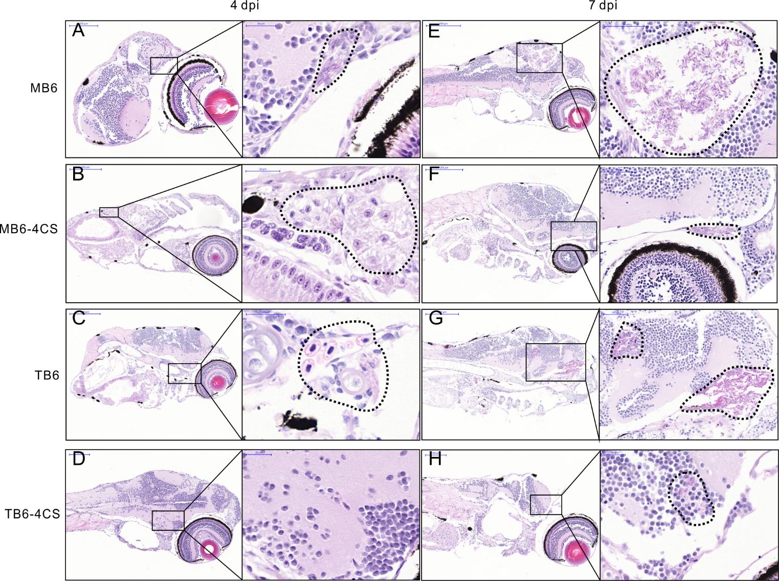
Fig. S7
Histological observation of holo- and apo-reduced-like WhiB6 in modulating replication and spread of Mm cells in zebrafish larvae. Related to Figure 6.
(A-H) Representative hematoxylin and eosin stains of zebrafish larvae 4 dpi (A-D) or 7 dpi (E-H) caudal infected with ~150 CFU of the indicated Mm strains. Areas enlarged are shown on the right of each image. Dotted circles delineate locations where Mm distributed, resulting in tissue damage and bacterial replication. Three fish per group were used for each time point.
Acknowledgments
This image is the copyrighted work of the attributed author or publisher, and
ZFIN has permission only to display this image to its users.
Additional permissions should be obtained from the applicable author or publisher of the image.
Full text @ Cell Rep.