
- Title
-
Smyd5 plays pivotal roles in both primitive and definitive hematopoiesis during zebrafish embryogenesis
- Authors
- Fujii, T., Tsunesumi, S.I., Sagara, H., Munakata, M., Hisaki, Y., Sekiya, T., Furukawa, Y., Sakamoto, K., Watanabe, S.
- Source
- Full text @ Sci. Rep.
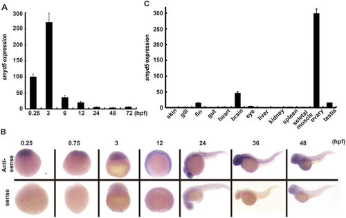
Expression patterns of smyd5 during zebrafish embryogenesis and in adult tissues. (A) qRT-PCR analysis was performed using smyd5 primer sets from RNAs extracted from zebrafish embryos at 0.25, 3, 6, 12, 24, 48, and 72 hpf. (B) In situ hybridisation of smyd5 at 0.25, 0.75, 3, 12, 24, 36, and 48 hpf. Lower and upper panels indicate smyd5 sense control and antisense probes, respectively. (C) qRT-PCR analysis of smyd5 in various adult tissues. Scale bar, 200 µm. |
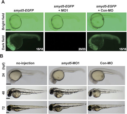
Effect of smyd5 knockdown by MO1 or MO2 in zebrafish embryos. (A) Suppression of smyd5 was examined at 24 hpf in embryos injected with smyd5-EGFP mRNA alone, smyd5-EGFP mRNA and MO1, and smyd5-EGFP mRNA and Con-MO. EGFP signals were examined by fluorescent microscopy (lower panel). Numbers on each panel indicate the number of embryos showing EGFP-positive embryos per total number of embryos. Morphogenesis of zebrafish embryos injected with MO1 (E) or Con-MO (F) at 24, 48, and 72 hpf. Embryos are depicted in the lateral view. Scale bar, 200 µm. |

Expression markers for heart and skeletal muscle in smyd5 morphants by WISH and electron microscopic analysis of heart, skeletal muscle, and slow muscle. WISH analysis of skeletal muscle and cardiac chamber markers at 12 (A), 24 (B), and 48 hpf (C). (D) Electron microscopic analysis of heat, skeletal muscle, and slow muscle at 48 hpf. (A) Expression of myf5, myod, myog, and gata5 in embryos injected with smyd5-MO1, control embryos injected with Con-MO, and those no-injection at 12 hpf. (B) Expression of myod, myog, and mck in embryos injected with smyd5-MO1, control embryos, and those no-injection at 24 hpf. (C) Expression of cmlc2 in morphants, control embryos, and those no-injection at 48 hpf. Embryos are shown in the dorsal view, anterior towards the left (A). Embryos are depicted in the lateral view (B) and in the frontal view, dorsal towards the left (C). (D) Electron micrographs of parasagittal sections through cardiac and somitic muscle cells of embryos injected with smyd5-MO1, and control embryos at 48 hpf. Numbers in the bottom of each panel indicate the number of embryos with the representative phenotype per the total number of examined embryos. Scale bar, 200 µm (black) and 1 µm (white). skm, skeletal muscle. EXPRESSION / LABELING:
|
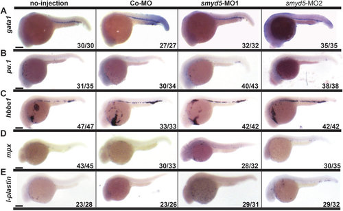
Expression of markers for primitive hematopoietic lineages in smyd5 morphants by WISH. Expression of gata1 (A) and pu.1 (B) in embryos injected with smyd5-MO1, smyd5-MO2, control embryos injected with Con-MO, and those no-injection at 24 hpf. Expression of hbbe1 (C), mpx (D), and l-plastin (E) in embryos injected with smyd5-MO1, smyd5-MO2, control embryos injected with Con-MO, and those no-injection at 26 (C) and 28 hpf (D,E). Numbers on each panel indicate the number of embryos showing the representative phenotype per the total number of embryos. Embryos are depicted in the lateral view. Scale bar, 200 µm. |
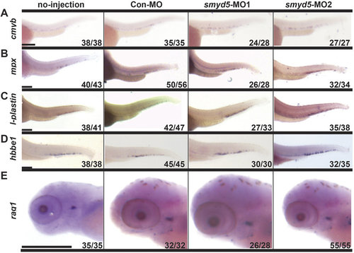
Expression of markers for definitive hematopoietic lineages in smyd5 morphants by WISH. (A) Expression of cmyb in embryos injected with smyd5-MO1, smyd5-MO2, control embryos injected with Con-MO, and those no-injection at 30 hpf. Expression of mpx (B) and l-plastin (C) in embryos injected with smyd5-MO1, smyd5-MO2, control embryos injected with Con MO, and those no-injection at 72 hpf. Expression of hbbe1 (D) and rag1 (E) in embryos injected with smyd5-MO1, smyd5-MO2, control embryos injected with Con-MO, and those no-injection at 96 hpf. The number of embryos with the representative phenotype per the total number of embryos is indicated in each panel. Embryos are depicted in the lateral view. Scale bar, 200 µm. EXPRESSION / LABELING:
PHENOTYPE:
|

CRISPR/Cas9 targeted mutation of smyd5 phenocopies morpholino knockdown. Identification of embryos with CRISPR/Cas9-mediated insertion and/or deletion (indel) mutations in smyd5 genomic region by heteroduplex mobility assay (HMA). Heteroduplex (whitelines) and homoduplex (asterisks) DNA band indicate the presence of indel mutant allele, and wild type allele, respectively. Five embryos (#1-#5) were injected with smyd5 guide RNA (gRNA) and Cas9 mRNA. We detected heteroduplex DNA band in #4 and #5 of embryos. Homoduplex DNA band was detected in #1, #2 and #3 of embryos, which has similar size with that observed in no-injected control. (B) Sequences of smyd5 mutations in #4 embryos. All sequences had indels near the smyd5 target site of gRNA, which is underlined. Deletions and insertions are indicated by dashe and lowercase red letters, respectively. The number of nucleotides deleted (-) and inserted (+) is indicated to the right with the detection number. (C,D) Whole mount in situ hybridization of smyd5-KO or control embryos. The genes involving primitive myelopoiesis in smyd5-KO F0 embryos were examined (C). Expression of pu.1 in smyd5-KO F0 embryos, and those no-injection at 24 hpf. Expression of mpx and l-plastinin in smyd5-KO F0 embryos and those no-injection at 28 hpf. Expression of the genes for definitive myelopoiesis in smyd5-KO F0 embryos was examined (D). Expression of cmyb in smyd5-KO F0 embryos, and those no-injection at 30 hpf. Expression of mpx and l-plastinin in smyd5-KO F0 embryos, and those no-injection at 72 hpf. Numbers on each panel indicate the number of embryos showing the representative phenotype per the total number of embryos. Embryos are depicted in the lateral view. Scale bar, 200 µm. EXPRESSION / LABELING:
PHENOTYPE:
|
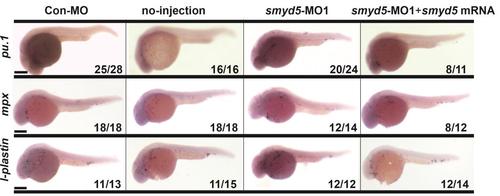
Phenotypic rescue experiments of primitive myelopoiesis in smyd5-knockdown embryos Expression of pu.1in embryos injected with Con-MO, those no-injection, smyd5-MO1, and smyd5-MO1 in combination with smyd5mRNA at 24 hpf. Expression of mpx andl-plastinin in embryos injected with Con-MO, those no-injection, smyd5-MO1, and smyd5-MO1 in combination with smyd5mRNA at 28 hpf. Numbers on each panel indicate the number of embryos showing the representative phenotype perthetotal number of embryos. Embryos are depicted in the lateral view. Scale bar, 200 µm. |

Phenotypic rescue experiments of definitive myelopoiesis in smyd5-knockdown embryos Expression of cmybin embryos injected with Con-MO, those no-injection, smyd5-MO1, and smyd5-MO1 in combination with smyd5mRNA at 30 hpf. Expression of mpx and l-plastinin in embryos injected with Con-MO, those no-injection, smyd5-MO1, and smyd5-MO1 in combination with smyd5mRNA at 72 hpf. Numbers on each panel indicate the number of embryos showing the representative phenotype perthetotal number of embryos. Embryos are depicted inthelateral view. Scale bar, 200 µm. |