
- Title
-
TWIST1 Integrates Endothelial Responses to Flow in Vascular Dysfunction and Atherosclerosis
- Authors
- Mahmoud, M., Kim, H.R., Xing, R., Hsiao, S., Mammmoto, A., Chen, J., Serbanovic-Canic, J., Feng, S., Bowden, N.P., Maguire, R., Ariaans, M., Francis, S., Weinberg, P.D., Van der Heiden, K., Jones, E.A., Chico, T.J., Ridger, V.C., Evans, P.C.
- Source
- Full text @ Circ. Res.
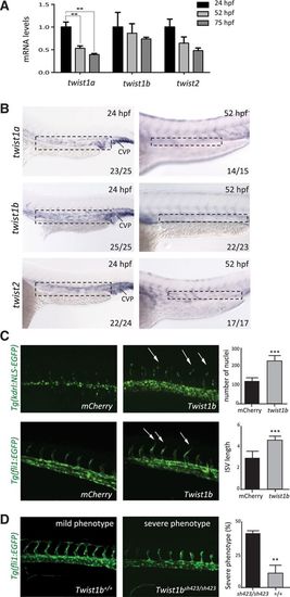
twist promoted intersegmental vessel sprouting in zebrafish embryos. A, The expression of twist1a, twist1b, and twist2 was studied at 24 to 75 hours post fertilization (hpf) by qPCR of the trunk and tail of embryos. Data were pooled from ne15 embryos studied in 3 independent experiments and mean values±SEs are shown. B, The expression of twist1a, twist1b, and twist2 was studied at 24 and 52 hpf by in situ hybridization. Data are representative of the majority of embryos analyzed (proportion indicated lower right of each part) and were closely similar in at least 3 independent experiments. Higher magnification insets are shown (marked in top). Bar=100 µm. C, Zebrafish embryos (wild-type, Tg(fli1:EGFP), or Tg(kdrl:NLS-EGFP)) were treated with twist1b mRNA (to enforce expression) or treated with mCherry mRNA as a control. They were studied at 24 to 27 hpf using confocal microscopy to visualize endothelial cell (EC) nuclei (Tg(kdrl:NLS-EGFP); top) or angiogenic sprouts (Tg(fli1:EGFP); bottom; arrows). Representative images are shown. Cell numbers and the length of intersegmental vessels (ISVs; third to fifth vessels in the field view) were quantified in multiple embryos, and mean values±SEM are shown (right). D, The twist1b coding sequence was mutated by introduction of a 4 bp deletion causing a frameshift and premature stop (mutant allele designated twist1bsh423). twist1bsh423/+ Tg(fli1:EGFP) fish were incrossed, and embryos were treated with a morpholino directed against twist1a. Sprouting of ISV was assessed at 34 hpf. Embryos were classified into those that displayed minimal sprouting (severe phenotype) and those with intermediate levels of sprouting (mild phenotype). Genotyping was subsequently performed, and the proportion of twist1b homozygous mutants (twist1bsh423/sh423) and twist1b homozygous wild-types (twist1b+/+) in the severe phenotype group was calculated (% indicated). Representative images are shown. Data were closely similar in 3 independent experiments. ***P<0.001 and **P<0.01 using a 1-way ANOVA (A) or unpaired t test (C and D). CVP indicates caudal vein plexus. EXPRESSION / LABELING:
PHENOTYPE:
|
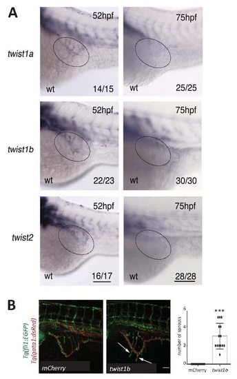
twist1 regulated sub-intestinal vein sprouting in embryos. (A) In situ hybridisation analysis of twist1a, twist1b or twist2 expression in embryos at 52 or 75 hpf. Scale bar, 100 µm. SIV region is indicated using a broken line. Data shown are representative of the majority of embryos analysed (proportion indicated lower right in each panel). (B) Embryos (Tg(fli1:EGFP;gata1:dsRed)) were treated with twist1b mRNA (to enforce expression) or with mCherry mRNA as a control. They were studied at 75 hpf (flow was established as evidenced by gata1-positive red blood cells). Confocal microscopy was used to visualise angiogenic sprouts (arrows). The number of angiogenic sprouts was quantified for multiple embryos and mean values +/- SEM are shown. Representative images are shown. Scale bar, 500 µm. *** p<0.001 using an unpaired t-test. |