
- Title
-
BNIP-H Recruits the Cholinergic Machinery to Neurite Terminals to Promote Acetylcholine Signaling and Neuritogenesis
- Authors
- Sun, J., Pan, C.Q., Chew, T.W., Liang, F., Burmeister, M., Low, B.C.
- Source
- Full text @ Dev. Cell
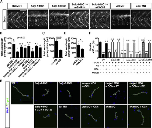
Knockdown of Bnip-h Causes Abnormal Axon Growth of Motor Neurons in Zebrafish through Cholinergic Signaling (A–D) The axon growth of motor neurons is regulated by Bnip-h, Acl and Chat. The axons of motor neurons were stained with anti-synaptotagmin-2 antibody (Znp-1) and imaged with confocal microscope. Images are maximum intensity projections of z stacks. Total neurite length per myotome (excluding myoseptum) was calculated. Five fishes for each condition were quantified. Error bars represent SD. p < 0.05; p < 0.001. Scale bar represents 100 µm. (E and F) Bnip-h regulates axon growth of primary motor neurons through muscarinic receptors and MAPK pathway. Primary motor neurons were isolated from 18 somite larvae, and treated with indicated chemicals for 48 hr. Quantification was done as in Figure 1B. n = 30–50 cells/category from three experiments. Error bars represent SD. p < 0.01; p < 0.001; N. S., not significant. Scale bar represents 50 µm. See also Figure S6. EXPRESSION / LABELING:
PHENOTYPE:
|
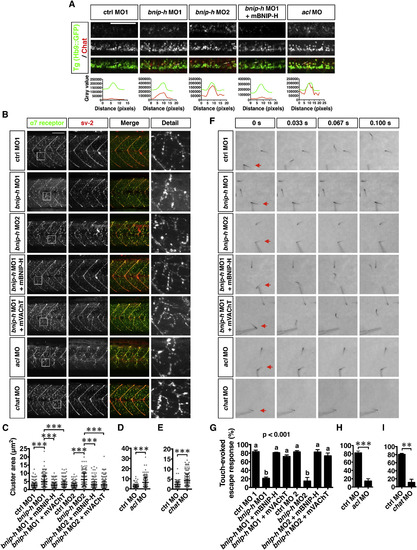
Knockdown of Bnip-h Impairs Cholinergic Signaling and Motility in Zebrafish (A) Chat molecules are mislocalized at the cell bodies of spinal motor neurons in bnip-h and acl morphants at 72 hpf. The larvae were stained with anti-Chat antibody and imaged with confocal microscope. Images are maximum intensity projections of z stacks. The fluorescent intensities of GFP (green) and Chat (red) across the lines in the cell bodies of motor neurons are shown in the lower image. (B–E) The Achr clustering is enlarged in bnip-h, acl, and chat morphants. The neuromuscular junctions were stained with α-Bungarotoxin (α7 receptor) and anti-synaptic vesicle glycoprotein 2A antibody (sv-2), and imaged with confocal microscope. Images are maximum intensity projections of z stacks. The size of Achr cluster was quantified. Error bars represent SD. p < 0.001. (F–I) The escape response to touching is blocked by Bnip-h, Acl or Chat knockdown. Escape responses of 72 hpf larvae were elicited by a gentle probe and recorded at rate of 300 frame/s. Percentages (%) of larvae with escape response are presented. n = 30–100 fishes/category from three experiments. Error bars represent SD. p < 0.01; p < 0.001. Scale bars represent 100 µm. See also Figure S7 and Movies S4 and >S5. EXPRESSION / LABELING:
PHENOTYPE:
|
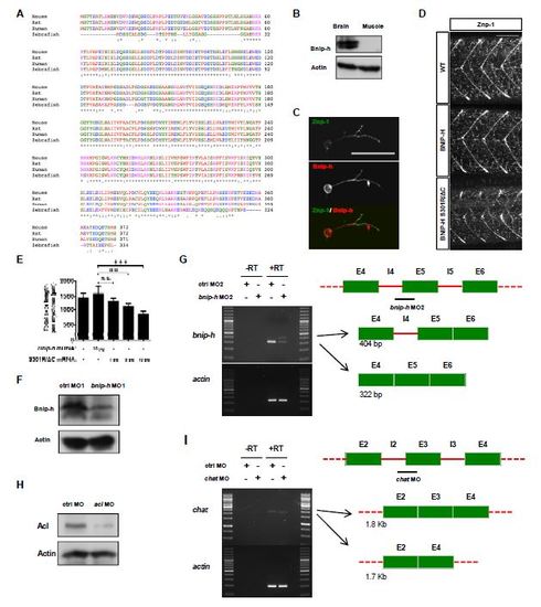
Knockdown of Bnip-h Causes Abnormal Axon Growth of Motor Neurons in Zebrafish through Cholinergic Signaling, Related to Figure 6 (A) Clustal W multiple sequence alignment of zebrafish, human, mouse and rat BNIP-H proteins (The asterisks indicate the conserved amino acids). (B) Bnip-h is expressed in brain but not in muscle of adult fish. (C) Bnip-h is expressed in zebrafish motor neurons. The primary motor neurons were isolated from zebrafish larvae at 24 hpf and cultured for additional 48 hours before fixation and staining. Scale bar, 50 µm. (D and E) BNIP-H S301R/ΔC mutant inhibits axon outgrowth of motor neurons in zebrafish larvae at 72 hpf. Quantification was done as in Figure 6B. Error bars represent SD. **p < 0.01; ***p < 0.001; N. S., not significant. Scale bar, 100 µm. (F) Bnip-h expression is reduced by the injection of bnip-h MO1 (translation-blocking morpholino) at 72 hpf. (G) The pre-mRNA splicing of bnip-h is disrupted by the injection of bnip-h MO2 (splice-site morpholino) at 72 hpf. Right: a schematic of wild type and abnormal bnip-h transcripts, based on the sequences of PCR products. (H) Acl expression is reduced by the injection of acl MO (translation-blocking morpholino) at 72 hpf. (I) The pre-mRNA splicing of chat is disrupted by the injection of chat MO (splice-site morpholino) at 72 hpf. Right: a schematic of wild type and abnormal chat transcripts, based on the sequences of PCR products. |

Knockdown of Bnip-h Impairs Cholinergic Signaling and Motility in Zebrafish, Related to Figure 7 (A) The structures of slow and fast muscles are disrupted by Bnip-h knockdown. The slow muscle fibers were stained with anti-myosin heavy chain 1/2/4/6 antibody (F59). The fast muscle fibers were stained with anti-myosin light chain 1/3f antibody (F310). Scale bar, 50 µm. (B, C and D) The escape response to tapping is blocked by Bnip-h, Acl or Chat knockdown. The larvae at 72hpf were placed in 10cm petri dishes, and the tapping stimulus was applied to one side of the dish with around 5 s intervals. The movement of larvae was recorded over 30 s. Percentage (%) of swimming larvae is presented. n = 30- 50 fishes/category from three experiments. Error bars represent SD. **p < 0.01. (E) The spontaneous movement of bnip-h morphants at 72 hpf is reduced. Similar amounts of larvae were placed into eppendorf tubes. The number of swimming larvae across the half depth of the water is quantified over 30 s. See also Movies S4-S5. EXPRESSION / LABELING:
PHENOTYPE:
|
Reprinted from Developmental Cell, 34(5), Sun, J., Pan, C.Q., Chew, T.W., Liang, F., Burmeister, M., Low, B.C., BNIP-H Recruits the Cholinergic Machinery to Neurite Terminals to Promote Acetylcholine Signaling and Neuritogenesis, 555-68, Copyright (2015) with permission from Elsevier. Full text @ Dev. Cell