
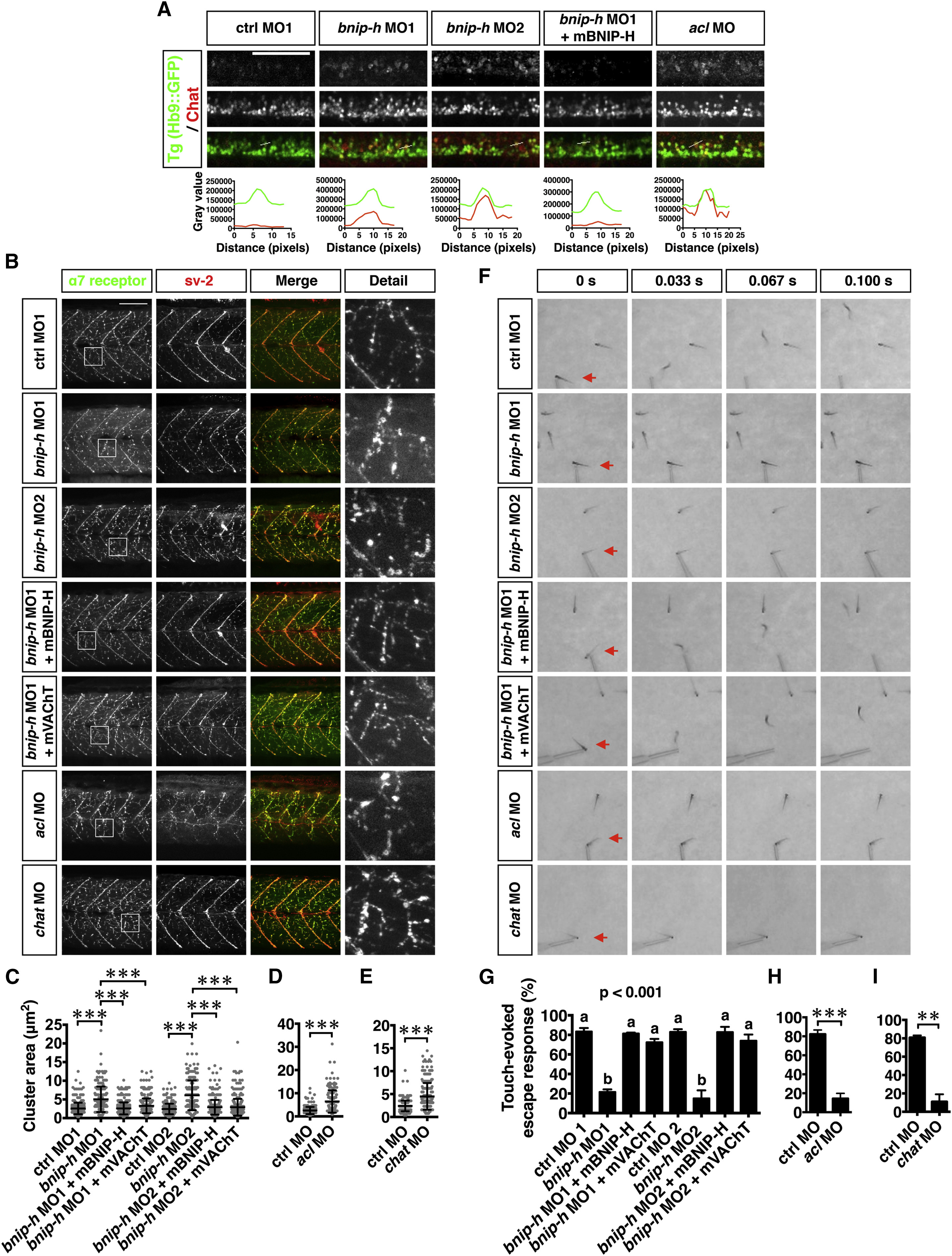
Fig. 7
|
Fig. 7
Knockdown of Bnip-h Impairs Cholinergic Signaling and Motility in Zebrafish
(A) Chat molecules are mislocalized at the cell bodies of spinal motor neurons in bnip-h and acl morphants at 72 hpf. The larvae were stained with anti-Chat antibody and imaged with confocal microscope. Images are maximum intensity projections of z stacks. The fluorescent intensities of GFP (green) and Chat (red) across the lines in the cell bodies of motor neurons are shown in the lower image.
(B–E) The Achr clustering is enlarged in bnip-h, acl, and chat morphants. The neuromuscular junctions were stained with α-Bungarotoxin (α7 receptor) and anti-synaptic vesicle glycoprotein 2A antibody (sv-2), and imaged with confocal microscope. Images are maximum intensity projections of z stacks. The size of Achr cluster was quantified. Error bars represent SD. p < 0.001.
(F–I) The escape response to touching is blocked by Bnip-h, Acl or Chat knockdown. Escape responses of 72 hpf larvae were elicited by a gentle probe and recorded at rate of 300 frame/s. Percentages (%) of larvae with escape response are presented. n = 30–100 fishes/category from three experiments. Error bars represent SD. p < 0.01; p < 0.001.
Scale bars represent 100 µm. See also Figure S7 and Movies S4 and >S5.
Reprinted from Developmental Cell, 34(5), Sun, J., Pan, C.Q., Chew, T.W., Liang, F., Burmeister, M., Low, B.C., BNIP-H Recruits the Cholinergic Machinery to Neurite Terminals to Promote Acetylcholine Signaling and Neuritogenesis, 555-68, Copyright (2015) with permission from Elsevier. Full text @ Dev. Cell