
- Title
-
Islet 1 specifies the identity of hypothalamic melanocortin neurons and is critical for normal food intake and adiposity in adulthood
- Authors
- Nasif, S., de Souza, F.S., González, L.E., Yamashita, M., Orquera, D.P., Low, M.J., Rubinstein, M.
- Source
- Full text @ Proc. Natl. Acad. Sci. USA
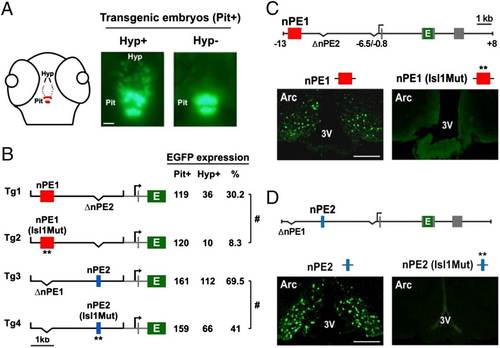
ISL1 binding sites in nPE1 and nPE2 are important for enhancer function. (A) Schematic of a 72-hpf zebrafish head showing the distribution of pomca-expressing cells (red dots) in the pituitary (Pit) and hypothalamus (Hyp). For transient reporter gene expression analyses, transgenic embryos showing strong pituitary EGFP expression (Pit+) at 72 hpf are selected and then sorted as expressing (Hyp+) or not expressing (Hyp) EGFP in the hypothalamus. (Scale bar, 50 µm.) (B) Expression analyses of transgenes driving EGFP under the control of zebrafish proximal pomca sequences (from 0.6 to +0.4 kb, in gray) and a neuronal mouse Pomc distal module carrying either wild-type or Isl1Mut enhancers nPE1 (red box) or nPE2 (blue box) in transgenic zebrafish. Seventy-two hours postfertilization, transgenic embryos were selected based on pituitary EGFP expression (Pit+), and the number of Pit+ embryos also showing EGFP+ neurons in the hypothalamus (Hyp+) was quantified. #P < 0.0001, Χ2 test. (C) Expression analysis of transgenic mice carrying nPE1 or nPE1 (Isl1Mut) sequences driving EGFP on coronal sections at the hypothalamic arcuate (Arc) level. (D) Expression analysis of transgenic mice carrying nPE2 or nPE2 (Isl1Mut) sequences driving EGFP on coronal sections at the hypothalamic arcuate (Arc) level. 3V, third ventricle. (Scale bar, 500 µm.) |
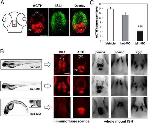
ISL1 regulates hypothalamic pomca expression in zebrafish. (A) Schematic of a 72-hpf zebrafish head in a horizontal plane showing the distribution of pomca-expressing cells in the pituitary and hypothalamus (red dots). The blue rectangle depicts the magnified area shown in the other panels. Double immunofluorescence analysis showing pomca (ACTH, red) and isl1 (green) coexpression in horizontal sections of 72-hpf zebrafish brain. (B, Left) Phenotypic characterization of 72-hpf Isl1-MO, het-MO, and vehicle-injected zebrafish. Isl1-MO larvae have reduced body size, curved down tail, and develop cardiac edema (indicated with an arrow in magnified Inset). (Right) Molecular characterization of the hypothalamus in Isl1-MO, het-MO, and vehicle-injected zebrafish embryos. Immunofluorescence (IF) analysis in horizontal brain sections of 72-hpf zebrafish shows that ISL1 knockdown completely eliminates pomca expression (ACTH) in the hypothalamus, but not in the pituitary. Whole-mount in situ hybridization (ISH) further confirms the lack of pomca expression in the brain and shows that other hypothalamic markers, pomcb and agrp, remain unchanged in Isl1-MO relative to control embryos. Hyp, hypothalamus; Pit, pituitary. (Scale bars, 50 µm.) (C) Quantification of the total number of ACTH+ neurons per embryo in Isl1-MO (n = 16)-, het-MO (n = 15)-, and vehicle (n = 28)-injected zebrafish. Bars correspond to mean + SEM. ***P < 0.001, Student’s t test vs. vehicle. EXPRESSION / LABELING:
PHENOTYPE:
|
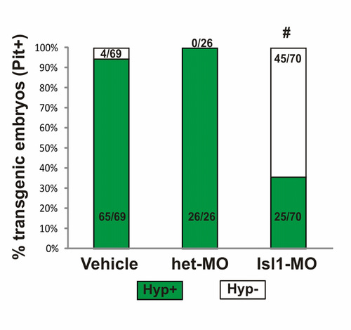
Analysis of EGFP expression in the hypothalamus of nPE1-nPE2zfpomca-EGFP transgenic zebrafish embryos injected with vehicle, het-MO or Isl1-MO. Transgenic embryos at 72 hpf are identified by pituitary EGFP expression (Pit+) and classified as expressing (Hyp+) or not expressing (Hyp-) EGFP in the hypothalamus. Isl1-MO injection significantly lowered the number of transgenic embryos showing hypothalamic EGFP expression. #p<1x10⁻¹⁰ Chi-square analysis. EXPRESSION / LABELING:
|