
- Title
-
Vegfd can compensate for loss of Vegfc in zebrafish facial lymphatic sprouting
- Authors
- Astin, J.W., Haggerty, M.J., Okuda, K.S., Le Guen, L., Misa, J.P., Tromp, A., Hogan, B.M., Crosier, K.E., Crosier, P.S.
- Source
- Full text @ Development
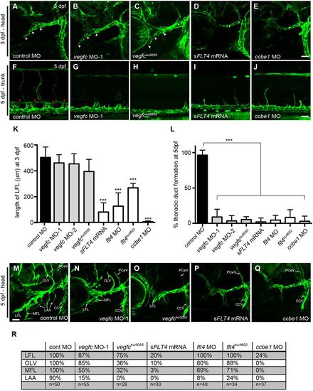
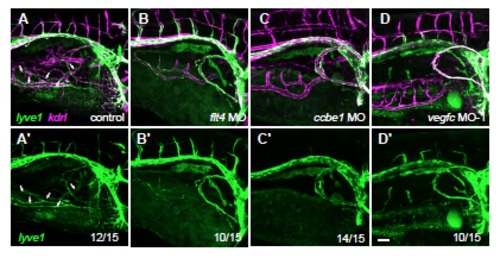
vegfc is not essential for zebrafish early facial lymphatic development. (A-J) Confocal images of the facial lymphatics in lyve1 embryos at 3dpf (A-E) or trunk lymphatics at 5 dpf (F-J) in control MO (A,F), vegfc MO-1 (B,G), vegfchu5055 (C,H), sFLT4 mRNA (D,I), and ccbe1 MO (E,J). Loss of Vegfc prevents formation of the TD (red asterisks), but the LFL forms normally at 3 dpf (white arrowheads) in vegfc morphant or vegfchu5055 mutant embryos. (K) Quantitation of LFL length at 3 dpf. (L) Quantitation of TD formation at 5 dpf. (M-Q) Confocal images of the facial lymphatics in lyve1 embryos at 5 dpf in control MO (M), vegfc MO-1 (N), vegfchu5055 (O), sFLT4 mRNA (P), and ccbe1 MO (Q). Knockdown of Flt4, Ccbe1 or Vegfc inhibits the correct development of the facial lymphatic network. Asterisk indicates the PHS. (R) The percentage formation of different facial lymphatic vessels at 5dpf. For flt4hu4602 and vegfchu5055 embryos, n=number of mutant embryos. CCV, common cardinal vein; PHS, primary head sinus; PCeV, posterior cerebral vein; LFL, lateral facial lymphatic; OLV, otolithic lymphatic vessel; MFL, medial facial lymphatic; LAA, lymphatic branchial arches. ***P<0.001, by a Mann–Whitney test versus control MO; error bars indicate s.d. Scale bars: 50 μm. EXPRESSION / LABELING:
PHENOTYPE:
|
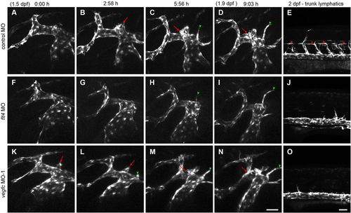
The facial lymphatic sprout forms normally in vegfc morphant embryos. (A-D,F-I,K-N) Stills from confocal time-lapse imaging of the facial lymphatic sprout in lyve1:egfp embryos from 1.5dpf to 1.9dpf (9:03h) with (E,J,O) a confocal image of the developing trunk lymphatics in the same embryo at 2dpf injected with either control MO (A-E; supplementary material Movie 1), flt4 MO (F-J; supplementary material Movie 2), or vegfc MO-1 (K-O; supplementary material Movie 3). Data are representative of three independent time-lapse experiments. The facial lymphatic sprout (red arrows) does not form in flt4 morphants but is present in vegfc morphant embryos that do not have parachordal lymphangioblasts (red arrowheads) in the trunk. The formation of the pectoral vein (green arrowheads) at approximately 6h into the experiment serves as a control to show that the embryos are developing normally. Scale bars: 50μm. |
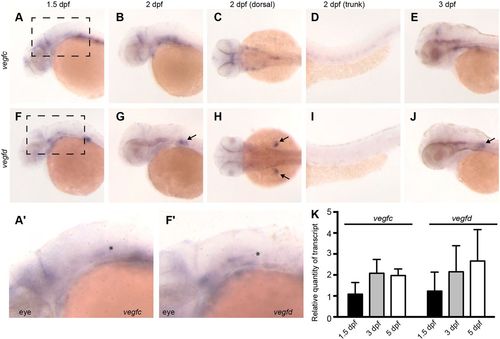
vegfd is expressed in the head and pectoral fin buds. Representative images of whole-mount in situ hybridisation showing the expression of vegfc (A-E) and of vegfd (F-J), in 1.5dpf (A,F), 2dpf (B-D,G-I) and 3dpf embryos (E,J). (C,H) The dorsal aspect of the head region at 2dpf and (D,I) lateral aspect of the trunk region at 2dpf. vegfd expression in the pectoral fin buds is marked with black arrows in G,H,J. (A2,F2) Magnified images of the boxed regions in A and F; the black asterisks show the approximate position of the FLS. (K) qPCR analysis of vegfc and vegfd mRNA levels from isolated heads of 1.5, 3 and 5dpf embryos. Data are shown relative to mRNA levels in 1.5dpf embryos. |
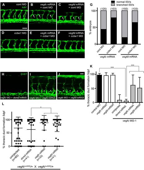
vegfd genetically interacts with ccbe1 and can compensate for loss of vegfc. (A-F) Confocal images of the trunk blood vasculature in 30hpf kdrl:egfp embryos injected with control MO (A-C), ccbe1 MO (D-F), 400pg vegfc mRNA (B,E) and 200pg vegfd mRNA (C,F). Injection of either vegfc or vegfd mRNA induced misguidance of the intersegmental blood vessels along the horizontal myoseptum (arrows). (G) The percentage of embryos with branched ISVs; numbers in brackets denote number of embryos scored. Silencing ccbe1 reduces the number of embryos with branched ISVs induced by injection of either vegfc or vegfd mRNA. (H-J) Confocal images of the trunk lymphatics in 5dpf lyve1:egfp embryos injected with vegfc MO-1+dsred mRNA (H), vegfc MO-1+400pg vegfc mRNA (I) and vegfc MO-1+200pg vegfd mRNA (J). Injection of either vegfc or vegfd mRNA can rescue the formation of the TD (asterisks) in vegfc morphant embryos. (K) Quantitation of TD formation at 5dpf. (L) Quantitation of TD formation at 5dpf in F1 progeny from an incross of vegfchu5055 heterozygous carriers. ***P<0.001, *P<0.05 by a Mann–Whitney test; error bars indicate s.d. Scale bars: 50μm. EXPRESSION / LABELING:
|
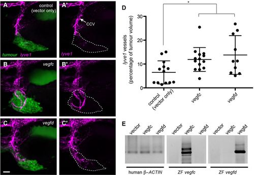
Expression of vegfd can induce ectopic lyve1-positive vessels. (A-C) Confocal images of human breast cancer cells labelled with Cell Tracker Green (MDA-MD-231) xenografted into lyve1:dsred embryos and imaged at 5dpf. (A) MDA-MD-231 cells transfected with vector only. (B) MDA-MD-231 cells transfected with zebrafish vegfc. (C) MDA-MD-231 cells transfected with zebrafish vegfd. (A2-C2) Confocal images of the lyve1:dsred vessels only. Cancer cells in green, lyve1 in magenta. Dashed lines indicate the boundary of the xenograft. The xenografts expressing either vegfc or vegfd are able to induce more ectopic lyve1-positive sprouts from the CCV than the control xenografts. (D) Quantitation of the tumour-induced lyve1-positive vessels, expressed as a percentage of tumour volume. (E) RT-PCR using RNA isolated from MDA-MB-231 cells transfected with vector only, vegfc or vegfd, with primers designed against human β-actin (ACTB), zebrafish vegfc or zebrafish vegfd. *P<0.05 by a Mann–Whitney test; error bars indicate s.d. Scale bar: 50μm. |
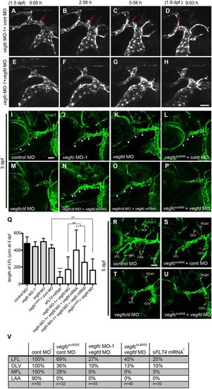
Knockdown of both vegfc and vegfd is required to prevent facial lymphatic development. (A-H) Stills from confocal time-lapse imaging of the facial lymphatic sprout in lyve1:egfp embryos from 1.5dpf to 1.9dpf (9:03h) injected with either vegfc MO-1 and control MO (A-D, supplementary material Movie 4) or vegfc MO-1 and vegfd MO (E-H, supplementary material Movie 5). Data are representative of three independent time-lapse experiments. The facial lymphatic sprout (red arrows) forms in vegfc MO-1 and control MO morphants but is not present in double-knockdown morphant embryos. The formation of the pectoral vein (green arrowheads) serves as a control to show that the embryos are developing normally. (I-P) Confocal images of the facial lymphatics in lyve1 embryos at 3dpf in control MO (I), vegfc MO-1 (J), vegfd MO (K), vegfchu5055+control MO (L), vegfc MO-1+vegfd MO (M), vegfc MO-1+vegfd MO+200pg vegfd mRNA (N), vegfc MO-1+vegfd MO+400pg vegfc mRNA (O) and vegfchu5055+vegfd MO (P). Silencing either vegfc or vegfd has no effect on early facial lymphatic development but double knockdown of both genes prevents the formation of the LFL at 3dpf (arrowheads). (Q) Quantitation of the length of the LFL vessel in 3dpf embryos (1data reproduced from Fig. 1 for comparison). (R-U) Confocal images of the facial lymphatics in lyve1 embryos at 5dpf in control MO (R), vegfchu5055+control MO (S), vegfc MO-1+vegfd MO (T) and vegfchu5055+vegfd MO (U). Asterisks indicates the PHS. (V) The percentage formation of different facial lymphatic vessels at 5dpf. (1data reproduced from Fig. 1 for comparison). For vegfchu5055 embryos, n=number of mutant embryos. Knockdown of both vegfc and vegfd results in a more severe defect in the development of the facial lymphatic network than knockdown of vegfc alone. CCV, common cardinal vein; PHS, primary head sinus; PCeV, posterior cerebral vein; LFL, lateral facial lymphatic; OLV, otolithic lymphatic vessel; MFL, medial facial lymphatic; LAA, lymphatic branchial arches. ***P<0.001, **P<0.01 by a Mann–Whitney test, unless indicated, significance was determined to control MO; error bars indicate s.d. Scale bars: 50μm. |
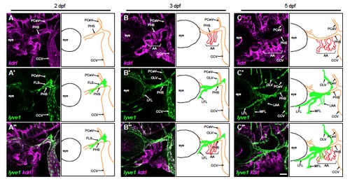
Zebrafish facial lymphatic development Confocal images of the developing facial lymphatics in lyve1:egfp;kdrl:rfp or lyve1:dsred;kdrl:egfp embryos at (A) 2 dpf (B) 3 dpf and (C) 5 dpf with lyve1 in green and kdrl in magenta with diagrams highlighting in red arteries that only express kdrl (AA), in orange veins that express both kdrl and lyve1 (PHS, CCV, and PCeV) and in green developing lymphatic vessels that only express lyve1 (FLS, LFL, MFL, OLV and LAA) at each stage. (A-C) Confocal images showing kdrl-expressing vessels. (A′-C′) Confocal images showing the lyve1-expressing vessels. (A′′-C′′) Confocal images of kdrl and lyve1 expression. CCV – common cardinal vein, PHS – primary head sinus, AA branchial arteries, PCeV – posterior cerebral vein, FLS – facial lymphatic sprout, LFL – lateral facial lymphatic, OLV – otolithic lymphatic vessel, MFL – medial facial lymphatic and LAA – lymphatic branchial arches. Scale bars: 50 μm. |
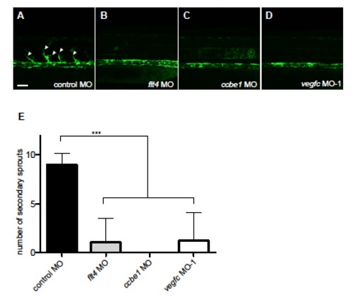
flt4, ccbe1 and vegfc are required for secondary sprouting from the PCV (A-D) Confocal images of 1.5 dpf lyve1:egfp embryos treated with (A) control MO (B) flt4 MO (C) ccbe1 MO (D) vegfc MO-1. Secondary sprouts from the PCV (arrowheads) are only present in embryos injected with control MO. (E) Quantitation of secondary sprout formation at 1.5 dpf in embryos injected with morpholinos. *** p<0.001, by a Mann-Whitney test to control; error bars indicate s.d. Scale bar: 50 μm. |
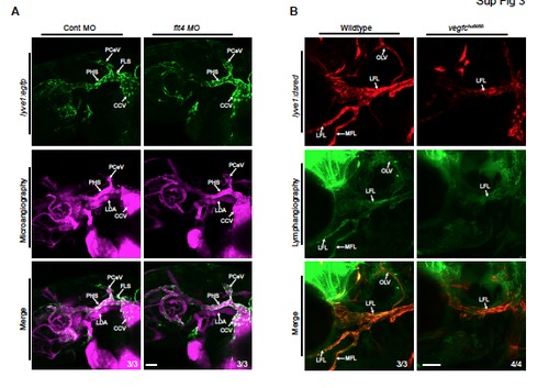
Microangiography and lymphangiography (A) Confocal images of 2 dpf lyve1:egfp embryos injected with fluorescent beads into the bloodstream to highlight blood flow. Vessels that express lyve1 (green) and contain blood flow (magenta) are veins (PHS, CCV and PCeV) while blood-containing vessels that do not express lyve1 are arteries (LDA). n=3 for control and flt4 morphants (B) Confocal images of 5 dpf lyve1:dsred embryos injected with fluorescein dextran to highlight functional lymphatics (lymphangiography). Data is representative of n=3 for wild type and n=4 for vegfchu5055. The vegfchu5055 mutant has a reduction in functional lymphatics at 5 dpf. CCV – common cardinal vein, PHS – primary head sinus, PCeV – posterior cerebral vein, LDA – lateral dorsal aorta, FLS – facial lymphatic sprout, LFL – lateral facial lymphatic, OLV – otolithic lymphatic vessel, MFL – medial facial lymphatic. Scale bars: 50 μm. |
flt4, ccbe1 and vegfc are required for development of the intestinal lymphatic network (A-D) Confocal images of the right intestinal lymphatics at 6 dpf in lyve1:dsred;kdrl:egfp embryos treated with (A) control MO (B) flt4 MO (C) ccbe1 MO (D) vegfc MO-1 with lyve1 in green and kdrl in magenta. (A′-D′) Confocal images of the lyve1:dsred vessels only. The right intestinal lymphatics are marked with arrows. flt4, ccbe1 and vegfc are all required for the formation of the intestinal lymphatic network. Scale bar: 50 μm. |
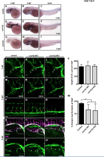
cxcr4a and cxcl12a are not required for development of the facial or intestinal lymphatics (A-I) Representative images of whole mount in situ hybridisation showing the expression of (A-C) cxcr4a (D-F) cxcl12a and (G-I) cxcl12b in (A,C,D,F,G) 2 dpf and (B,E,H,I) 3 dpf embryos showing that cxcr4a, cxcl12a and cxcl12b are expressed in the head. (J-O) Confocal images of the facial lymphatics in lyve1 embryos at (J-L) 3 dpf (M-O) 5 dpf (PR) trunk lymphatics at 5 dpf (S-U) intestinal lymphatics at 6 dpf in lyve1:dsred;kdrl:egfp embryos injected with (J,M,P,S) control MO (K,N,Q,T) cxcr4a MO or (L,O,R,U) cxcl12a MO. (S′-U′) Confocal images of the lyve1:dsred vessels only. lyve1 in green and kdrl in magenta. The LFL (white arrowheads) and right intestinal lymphatics (white arrows) form normally in cxcr4a or cxcl12a morphant embryos but the development of the thoracic duct (red asterisks) is impaired. The PHS is marked with a white asterisk. (V) Quantitation of the length of the lateral facial lymphatic (LFL) vessel in 3 dpf embryos. (W) Quantitation of the percentage thoracic duct formation in 5 dpf embryos. ** p<0.01, by a Mann-Whitney test to control; error bars indicate s.d. Scale bars: 50 μm. |
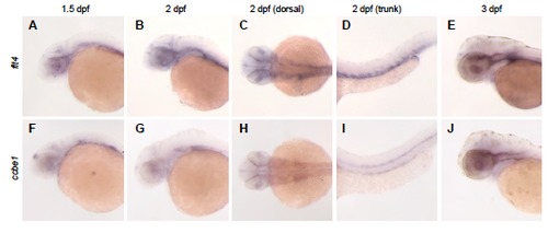
Expression of flt4 and ccbe1 Representative images of whole mount in situ hybridisation showing the expression of (A-E) flt4, and (F-J) ccbe1 in (A,F,) 1.5 dpf, (B-D,G-I) 2 dpf and (E,J) 3 dpf embryos. Development | Supplementary Material (C,H) The dorsal aspect of the head region at 2 dpf and (D,I,) lateral aspect of the trunk region at 2 dpf. EXPRESSION / LABELING:
|
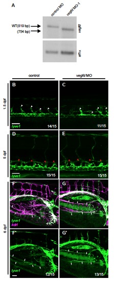
vegfd is not essential for development of the trunk or intestinal lymphatic networks (A) RT-PCR using RNA isolated from 1 dpf embryos injected with either control MO or 1 ρmole of vegfd MO using primers against vegfd (top panel) or ef1α (bottom panel). The vegfd MO causes aberrant splicing of the vegfd mRNA, resulting in deletion of 65 bp. (B-E) Confocal images of the trunk lymphatics in lyve1:egfp embryos at (B,C) 1.5 dpf (D,E) 5 dpf and (F,G) intestinal lymphatics at 6 dpf in lyve1:dsred;kdrl:egfp embryos injected with (B,D,F) control MO or (C,E,G) vegfd MO. (F′,G′) Confocal images of the lyve1:dsred vessels only. lyve1 in green and kdrl in magenta. vegfd morphants display normal secondary sprouting from the PCV (white arrowheads), normal thoracic duct formation (red asterisks), and normal intestinal lymphatic formation (white arrows). Scale bars: 50 μm. |