
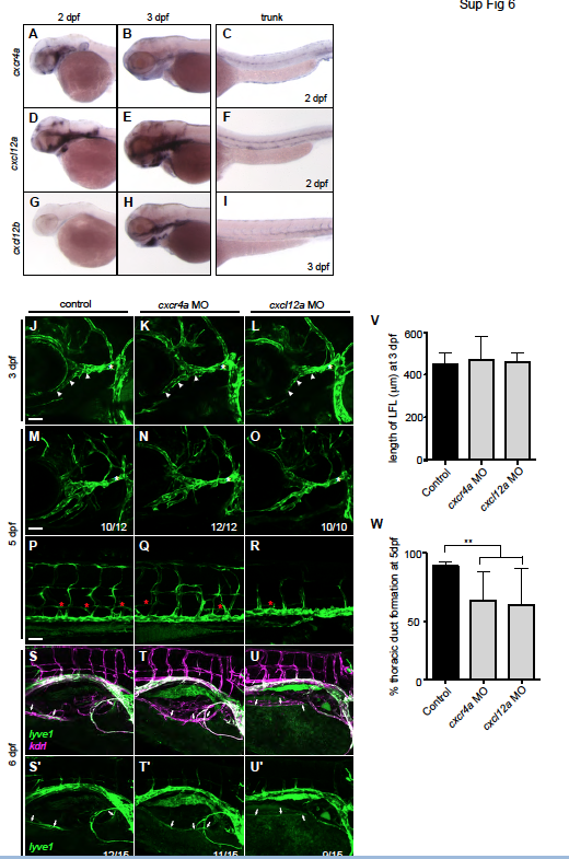
Fig. S6
|
Fig. S6
cxcr4a and cxcl12a are not required for development of the facial or intestinal lymphatics
(A-I) Representative images of whole mount in situ hybridisation showing the expression of (A-C) cxcr4a (D-F) cxcl12a and (G-I) cxcl12b in (A,C,D,F,G) 2 dpf and (B,E,H,I) 3 dpf embryos showing that cxcr4a, cxcl12a and cxcl12b are expressed in the head. (J-O) Confocal images of the facial lymphatics in lyve1 embryos at (J-L) 3 dpf (M-O) 5 dpf (PR) trunk lymphatics at 5 dpf (S-U) intestinal lymphatics at 6 dpf in lyve1:dsred;kdrl:egfp embryos injected with (J,M,P,S) control MO (K,N,Q,T) cxcr4a MO or (L,O,R,U) cxcl12a MO. (S′-U′) Confocal images of the lyve1:dsred vessels only. lyve1 in green and kdrl in magenta. The LFL (white arrowheads) and right intestinal lymphatics (white arrows) form normally in cxcr4a or cxcl12a morphant embryos but the development of the thoracic duct (red asterisks) is impaired. The PHS is marked with a white asterisk. (V) Quantitation of the length of the lateral facial lymphatic (LFL) vessel in 3 dpf embryos. (W) Quantitation of the percentage thoracic duct formation in 5 dpf embryos. ** p<0.01, by a Mann-Whitney test to control; error bars indicate s.d. Scale bars: 50 μm.