
|
Fig. 5
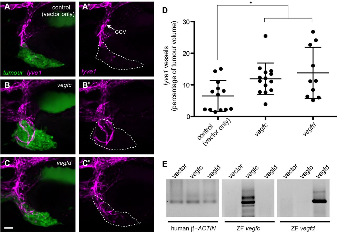
Expression of vegfd can induce ectopic lyve1-positive vessels. (A-C) Confocal images of human breast cancer cells labelled with Cell Tracker Green (MDA-MD-231) xenografted into lyve1:dsred embryos and imaged at 5dpf. (A) MDA-MD-231 cells transfected with vector only. (B) MDA-MD-231 cells transfected with zebrafish vegfc. (C) MDA-MD-231 cells transfected with zebrafish vegfd. (A2-C2) Confocal images of the lyve1:dsred vessels only. Cancer cells in green, lyve1 in magenta. Dashed lines indicate the boundary of the xenograft. The xenografts expressing either vegfc or vegfd are able to induce more ectopic lyve1-positive sprouts from the CCV than the control xenografts. (D) Quantitation of the tumour-induced lyve1-positive vessels, expressed as a percentage of tumour volume. (E) RT-PCR using RNA isolated from MDA-MB-231 cells transfected with vector only, vegfc or vegfd, with primers designed against human β-actin (ACTB), zebrafish vegfc or zebrafish vegfd. *P<0.05 by a Mann–Whitney test; error bars indicate s.d. Scale bar: 50μm.