
- Title
-
Ginger Stimulates Hematopoiesis via Bmp Pathway in Zebrafish
- Authors
- Ferri-Lagneau, K.F., Moshal, K.S., Grimes, M., Zahora, B., Lv, L., Sang, S., and Leung, T.
- Source
- Full text @ PLoS One
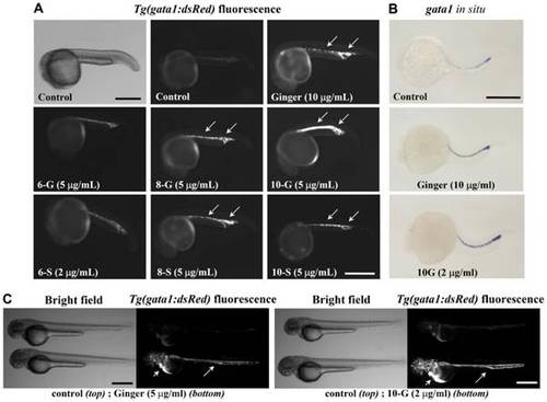
Ginger extract and its purified phenolic compounds promote Tg(gata1:dsRed) fluorescence and gata1 mRNA expression. (A) Bright field (top left) and Tg(gata1:dsRed) fluorescence of zebrafish embryos at about 22 hpf, before the onset of circulation (anterior to the left). Exposure to ginger extract or its compounds 8-gingerol (8-G), 10-gingerol (10-G), 8-shogaol (8-S) and 10-shogaol (10-S) promoted Tg(gata1:dsRed) fluorescent erythroid cell development in the ICM and PBI (arrows), as compared to control embryos. N = 35 embryos per group. In this panel, we show an embryo treated with a lower concentration of 6-S (2 μg/ml) as this compound was toxic at higher doses. Scale bar = 400 μm. (B) Whole-mount in situ hybridization of ginger or 10-G treated embryos (8 hpf to 21 hpf exposure) revealed increased expression of gata1 transcript at 22 hpf. N = 50 embryos per group. Scale bar = 350 μm. (C) At 48 hpf, control embryos at the top; ginger or 10-G treated embryos at the bottom. Scale bar = 500 μm. Fluorescent erythrocytes circulating in the axial vasculature (arrows) and in the pericardial space (arrow heads). |
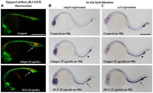
Ginger/10-G treatment increases hematopoietic progenitor markers expression. Zebrafish embryos were treated with ginger or 10-G from 9 to 21 hpf. (A) Tg(gata1:dsRed) for erythrocyte and Tg(flk1:GFP) for blood vessels, double-fluorescent overlay pictures of embryos at 22 hpf after exposure to ginger or 10-G. Hypertrophy of the PBI vascular plexus in Tg(flk1:GFP) after ginger or 10-G treatment, with Tg(gata1:dsRed) red fluorescent erythrocytes accumulated inside the honeycomb-like vasculature (arrows). Scale bars = 500 μm. Whole-mount in situ hybridization of c-myb (B) and scl (C) in zebrafish embryos at 22 hpf. Both hematopoietic progenitor markers were up-regulated in primitive hematopoietic tissues (ICM+PBI) upon ginger (arrow head) or 10-G (arrow) exposure. Scale bar = 350 μm. |
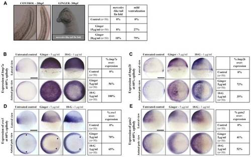
Ginger/10-G treatment during gastrulation promotes bmp2b/7a and Bmp target gene expression in zebrafish embryos. (A) Treatment of late gastrulae with ginger at 15 or 20 μg/ml induces the mercedes mutant-like phenotype (partial duplication of the tail fin) at 1 dpf in 8% or 10% of the treated embryos, respectively. Thus, the zebrafish embryos exposed to ginger extract mimic the phenotype of the ogon mutant, which has a mutation in sizzled, a bmp suppressor gene, at 1 dpf. (B) bmp7a expression was strongly increased and extended to the entire blastoderm at 60% epiboly, following short-term exposure to ginger (5 μg/ml) or 10-G (1 μg/ml) from sphere (4 hpf) to 60% epiboly (7 hpf) stages. (C) Up-regulation and extension of the expression domain were observed for bmp2b at 60% epiboly. (D–E) Accordingly, BMP target genes were up-regulated after ginger/10G treatment from the sphere stage (4 hpf) to 7 hpf, as illustrated by enhanced eve1 extended towards the dorsal side (arrow heads), a ventral mesoderm marker (D), and gata2, a non-neural ectoderm marker (E), in zebrafish embryos at 60% epiboly. Pictures on left panels show gastrulae, dorsal side to the right (B–E) and statistics tables (right panels) are representative of three independent experiments. N = number of embryos per group. Scale bars = 250 μm. |
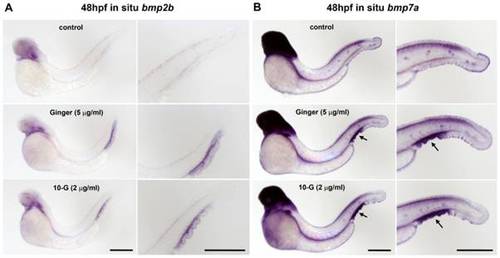
Ginger/10-G treatment after gastrulation promotes bmp2b/7a in the developing caudal hematopoietic tissue. (A–B) Zebrafish embryos were treated with ginger (5 µg/ml) or 10-G (2 μg/ml) from 10 to 48 hpf, followed by whole-mount in situ hybridization of bmp2b (A) and bmp7a (B). Both bmp2b and bmp7a were up-regulated locally in the CHT (and underlying fin) upon ginger or 10-G exposure (whereas they are not expressed in the CHT of control embryos at 48 hpf). Scale bars = 700 μm. |
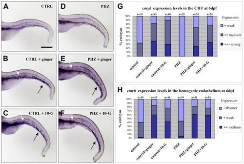
Effect of ginger and 10-G treatments on c-myb expression in zebrafish embryos at 6 dpf. (A–C) Ginger (B) or 10-G (C) treatment from 2 dpf to 6 dpf promotes cmyb expression in the CHT (black arrows) and the hemogenic endothelium (white arrows) along the ventral wall of the dorsal aorta (AGM equivalent) in the trunk and tail regions of normal embryos. (D–F) In phenylhydrazine-induced anemic embryos, ginger (E) or 10-G (F) treatment similarly promotes cmyb expression both in the CHT (black arrows) and the hemogenic endothelium (white arrows). (G–H) Graphical representation of the percentage of embryos showing cmyb expression in the CHT (G) and in the hemogenic endothelium (H). CTRL: control; PHZ: phenylhydrazine; n = number of embryos. Scale bar = 500 μm. |
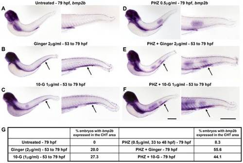
Over-expression of bmp2b specifically localized in the CHT area at 79 hpf upon ginger or 10-G exposure in normal and in anemic zebrafish embryos. Whole-mount in situ hybridization of bmp2b. (A–C, left) Normal non-anemic control embryos or embryos treated with ginger/10-G. (D–F, right) Anemic control embryos or anemic embryos treated with ginger/10-G. Anemic groups were treated with 0.5 μM PHZ from 33 to 48 hpf. Embryos express bmp2b in the CHT region (arrows) following exposure to ginger (B, E) or 10-G (C, F). (G) A table shows the percentage of embryos with bmp2b expression in the CHT area at 79 hpf. Scale bars = 420 μm. |
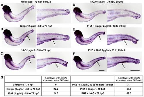
Over-expression of bmp7a specifically localized in the CHT region at 79 hpf upon ginger or 10-G exposure in normal and in anemic zebrafish embryos. Whole-mount in situ hybridization of bmp7a. (A–C, left) Normal non-anemic control embryos or embryos treated with ginger/10-G. (D–F, right) Anemic control embryos or anemic embryos treated with ginger/10-G. Anemic group were treated with 0.5 μM PHZ from 33 to 48 hpf. Embryos express bmp7a in the CHT area (arrows) following exposure to ginger (B, E) or 10-G (C, F). (G) A table shows the percentage of embryos with bmp7a expression in the CHT region at 79 hpf. Scale bars = 420 μm. |
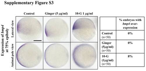
bmp4 expression in late gastrulae exposed to ginger/10-G. Whole mount in situ analysis of bmp expression after treatment with ginger/10-G. The bmp4 expression pattern at 75% epiboly was not affected by short-term treatment with ginger/10-G from sphere (4 hpf) to 75% epiboly (8 hpf) stages during early development. Embryos are oriented with the dorsal side to the right. Scale bar = 250 μm. |
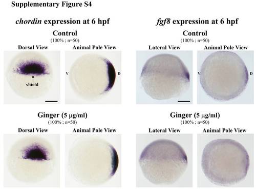
Ginger treatment of zebrafish embryos does not affect the expression of chd and fgf8. Whole mount in situ of chordin (chd) and fgf8 after treatment with ginger/10-G. Normal expression patterns of both chd at the dorsal margin (left panel) and fgf8 at the dorsal and ventral margins (right panel) at the shield stage after treatment with ginger/10-G. Lateral and animal views of representative embryos, with the dorsal side (D) to the right, ventral (V) to the left. Scale bars = 200 μm. |
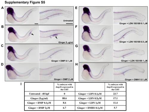
Bmp antagonists, that inhibit the canonical BMP-Smad signaling pathway, suppress the ginger-induced bmp2b expression in the region of the developing CHT. Whole mount in situ hybridization of bmp2b expression in zebrafish embryos after treatment with Bmp inhibitors and/or ginger/10-G from 10 to 48 hpf. (A) A control embryo. (B) Zebrafish embryos treated with ginger (5 µg/ml). (C–D) Ginger (5 μg/ml) and Dorsomorphin/DMP, 0.1 and 2 μM. (E–G) Ginger (5 μg/ml) and LDN193189, 0.1, 0.5 and 1 μM. (H) Ginger (5 μg/ml) and DMH1, 0.1 μM. (I) Analyses of bmp2b expression localized in the CHT area (table). Scale bars = 300 μm. |
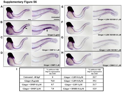
Bmp/Smad signaling antagonists inhibit the ginger-induced bmp7a expression in the area of the developing CHT. Whole-mount in situ hybridization of bmp7a in zebrafish embryos, after treatment with Bmp inhibitors and/or ginger/10-G from 10 to 48 hpf. (A) A control embryo. (B) Zebrafish embryos treated with ginger (5 μg/ml). (C–D) Ginger (5 μg/ml) and dorsomorphin/DMP, 0.1 and 2 μM. (E–G) Ginger (5 μg/ml) and LDN193189, 0.1, 0.5 and 1 μM. (H) Ginger (5 μg/ml) and DMH1, 0.1 μM. (I) Analyses of bmp7a expression localized in the CHT region (table). Scale bars = 300 μm. |
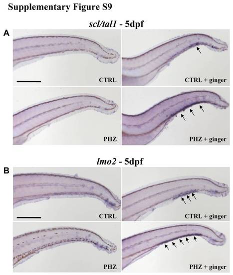
Up-regulation of hematopoietic progenitor markers scl/tal1 and lmo2 in the CHT at 5 dpf following ginger treatment in anemic zebrafish embryos. (A–B) Whole-mount in situ hybridization of scl/tal1 (A) and lmo2 (B) showing over-expression of these hematopoietic progenitor markers in the CHT (arrows) of anemic embryos treated with ginger extract. Scale bars = 500 μm. |