
- Title
-
Bmp and Nodal Independently Regulate lefty1 Expression to Maintain Unilateral Nodal Activity during Left-Right Axis Specification in Zebrafish
- Authors
- Smith, K.A., Noël, E., Thurlings, I., Rehmann, H., Chocron, S., and Bakkers, J.
- Source
- Full text @ PLoS Genet.
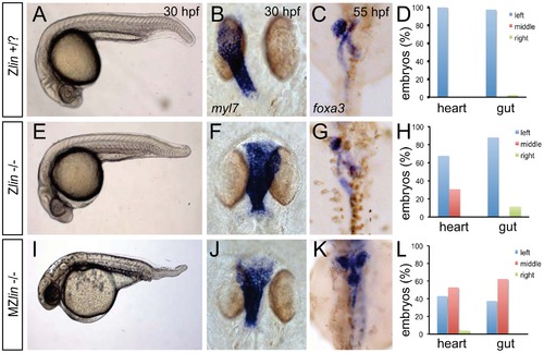
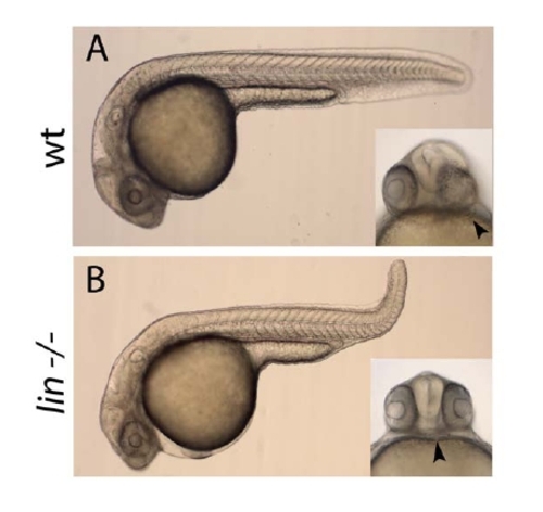
Dorsoventral and laterality defects in zygotic and maternal zygotic lin mutant embryos. (A–D) Wild-type zygotic lin siblings with normal ventral tail fin (A), left-positioned heart tube (B) and normal organ situs with liver on the left, pancreas on the right and left looped gut tube (C). (D) Quantification of heart position (n = 14) and direction of gut looping (n = 40). (E–H) Zygotic lin (Zlin) mutant embryos displayed a mild reduction of the ventral tail fin (n = 100/108) (E). In addition, in almost 30% of Zlin mutant embryos, the heart tube was positioned at the midline (F). Gut laterality was unaffected in Zlin mutant embryos (G). Quantification of heart position (n = 108) and direction of gut looping (n = 100). (I–L) Maternal zygotic lin (MZlin) mutant embryos derived from a cross of a homozygous lin mutant female and male showing the more severe posterior truncation (I) compared to a Zlin mutant embryo (E). In addition, most MZlin mutant embryos displayed a laterality defect in the heart (J), liver (bilateral, K) and in looping of the gut (K). (L) Quantification of heart positioning (n = 151) and direction of gut looping (n = 16). |
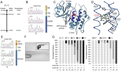
Genetic variations found in zebrafish bmpr1aa and bmpr1ab genes. (A) The lin mutation was mapped to a region on chromosome 13 that includes the bmpr1aa gene. (B) T>A basepair change that was found in all lin mutant embryos results in a Leu to Arg change at position 337 (L337R). (C) Crystal structure of human BMPR1B. The kinase domain from the human BMPR1B with the kinase inhibitor LDN-193189 (ball-and-stick representation) bound to the ATP binding site (pdb entry 3MDY). Leu 312 (corresponding to Leu 337 in fish) is shown in red. Structural elements providing residues to the hydrophobic core surrounding Leu 312 are highlighted in dark blue. (D) Detailed view of the hydrophobic core surrounding Leu 312 (in red). Black labels refer to the structure of human BMPR1A, the corresponding residues in fish are indicated by grey italic labels. Consequences of the L312R mutation are analyzed by replacing the leucine side chain in the structure model with arginine, of which five typical rotamers are shown (yellow to green). All rotamers cause serious clashes with surrounding residue, which are highly conserved in fish. (E) C>A basepair change in the bmpr1ab gene that results in a premature stop codon at position 84 in the extracellular domain of the receptor. (F) A MZbmpr1ab mutant embryo at 2 dpf with no obvious phenotypes in the heart or tail region (magnified). (G) Bmpr1a dose-dependent effect on dorsoventral and left-right patterning. Embryos derived from an incross of bmpr1aa+/-bmpr1ab+/- double carrier fish was analyzed and quantified for the dorsoventral phenotypes (classified as C1 (mild) to C4 (strong)) and position of the heart (left or midline) if present. n/a, not applicable since no heart tissue was present. PHENOTYPE:
|
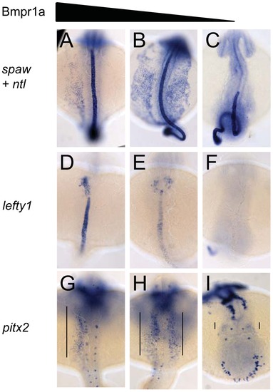
Dose-dependent effect of Bmpr1a on the expression of laterality genes. In situ hybridisation at 18-somites for spaw (in LPM) and no tail (ntl) (in midline) (A–C), lefty1 at 23-somites (heart field and midline) (D–F) and pitx2 at 23-somites (in LPM) (G–I). (A,D,G) Embryos selected for normal ventral tail fin or C1 dorsalization (genotypes: bmpr1aa +/+ or +/- bmpr1ab +/+ or +/- or -/-). (B,E,H) Embryos selected for C3 dorsalization (genotype bmpr1aa-/-;bmp1ab+/-). (C,F,I) Embryos selected for C4 dorsalization (genotype bmpr1aa-/-;bmpr1ab-/-). All embryos are shown as dorsal views with anterior to the top and left to the left. Number of embryos examined is presented in Table 2. EXPRESSION / LABELING:
PHENOTYPE:
|
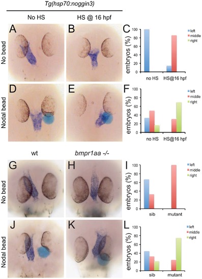
Rescue of Bmp-related cardiac laterality defects by Nodal beads. In situ hybridisation for myl7 to highlight the position of the linear heart tube at 30 hpf. Tg(hsp70l:nog3) embryos with no heat-shock (A,D) or heat-shocked at 16 hpf (B,E). Beads (blue) preincubated with recombinant Nodal protein placed in the right ALPM of non-heat-shocked (D) or heat-shocked (E) Tg(hsp70l:nog3) embryos at 17–18 hpf. Control siblings (G,J) or MZbmpr1aa mutant embryos (H,K). Beads (blue) preincubated with Nodal protein placed in the right ALPM of siblings (J) or MZbmpr1aa mutant embryos (K). Position of the inflow pole of the linear heart tube was determined for embryos without a Nodal bead (C) and for embryos in which a Nodal bead was placed on the right side (F). Embryos are shown as dorsal views with anterior to the top and left to the left. |
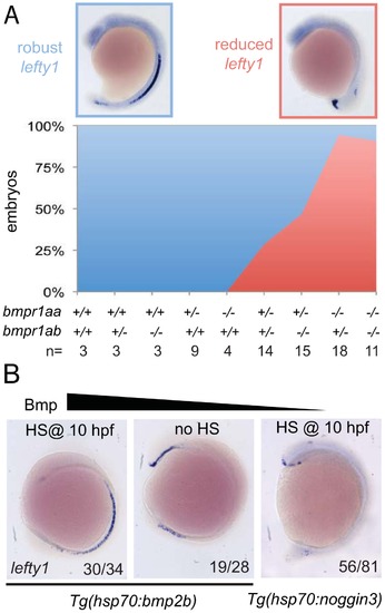
Bmp via Bmpr1a regulates lefty1 expression in the midline. (A) In situ hybridisation for lefty1 at 15-somites on embryos from an incross of bmpr1aa+/-;bmpr1ab+/- double carrier fish. Embryos were analysed for lefty1 expression and classified as robust (blue boxed panel) or reduced (red boxed panel) expression after which the embryos were genotyped. Quantification of the results is shown in the stacked area graph (blue, robust lefty1; red reduced lefty1). (B) In situ hybridisation for lefty1 at 10-somite stage. Embryos shown are Tg(hsp70l:bmp2b) embryos either heat-shocked at 10 hpf to induce bmp2b expression (left panel) or without heat-shock (middle panel) and Tg(hsp70l:nog3) embryos heat-shocked at 10 hpf to inhibit Bmp signalling (right panel). Lateral view of 10-somite stage embryos with dorsal to the right and anterior up. EXPRESSION / LABELING:
|
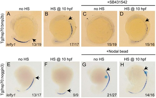
Bmp and Nodal induce lefty1 independently. (A–D) Tg(hsp70l:bmp2b) embryos were left at 28°C (A,C) or heat-shocked at 10 hpf to induce bmp2b expression (B,D). A subset of embryos were incubated in the presence of the Nodal inhibitor SB431542 directly after the heat-shock. Embryos were analysed by in situ hybridisation for lefty1 expression at 15-somites. (E–H) Tg(hsp70l:nog3) embryos were left at 28°C (E,G) or heat-shocked at 10 hpf to induce noggin3 expression (F,H). In a subset of embryos a bead preincubated with recombinant Nodal was placed in the ALPM. Embryos were analysed by in situ hybridisation for lefty1 expression at 18-somites. All embryos are shown as lateral views with dorsal to the right and anterior to the top. Arrows point to most anterior lefty1 expression domain. Numbers in lower right represents the number of embryos that displayed the phenotype represented in the panels. EXPRESSION / LABELING:
|
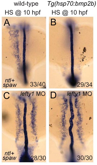
Lefty1 is required for Bmp induced repression of spaw. In situ hybridisation of spaw (in LPM) and ntl (in midline) at 18-somites. Wild-type (A,C) or Tg(hsp70l:bmp2b) (B,D) embryos were heat-shocked at 10 hpf to induce bmp2b expression (B,D). Ectopic expression of bmp2b resulted in the loss of spaw expression in the LPM (B). A subset of embryos were injected with a lefty1 MO (C,D), which resulted in bilateral spaw expression even in the presence of ectopic bmp2b (D). Embryos are shown as dorsal views with anterior to the top and left to the left. EXPRESSION / LABELING:
|
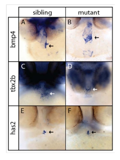
Formation of the cardiac atrioventricular canal is unaffected in bmpr1aa mutant embryos. (A,B) In situ hybridization for bmp4 in the heart of wild-type and bmpr1aa mutant embryos at 48 hpf. Bmp4 is expressed in the inflow region, atrioventricular (AV) canal (arrow) and outflow region of the heart. Although cardiac looping was affected in bmpr1aa mutant embryos, expression of bmp4 was unaffected. (C,D) In situ hybridization for tbx2b, which was expressed in the AV canal in wild-type siblings (C) and bmpr1aa mutant embryos (D). (E,F) In situ hybridization for has2, which was expressed in the endocardial cushion cells that will form the AV valves. Has2 expression was unaffected in bmpr1aa mutant embryos (F) compared to its wild-type siblings (E). |
Cilia rotation in Kupffer′s vesicle of Zlin mutant is unaffected. Brightfield images of the heart of wt and zygotic lin mutants after imaging cilia in the KV. Zygotic lin mutants display defects in positioning of the heart, however cilia motility in the KV is unaffected (Videos S1 and S2), demonstrating cilia-independent heart defects. |
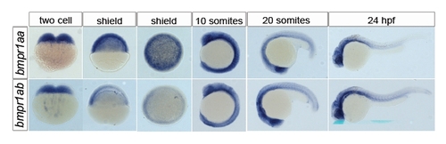
Expression of bmpr1aa and bmpr1ab. In situ hybridization for bmpr1aa (upper row) and bmpr1ab (lower row) at the indicated stages from 2-cells up to 24 hpf. Both maternal bmpr1aa mRNA and bmpr1ab mRNA was detected at the 2-cell stage. mRNA for both Bmp receptors was detected at the various developmental stages up to 24 hpf. Whilst expression of both Bmp receptors was distributed ubiquitously up to the 10-somite stage, it became progressively more intense in anterior structures at the 20-somite stages and later. |
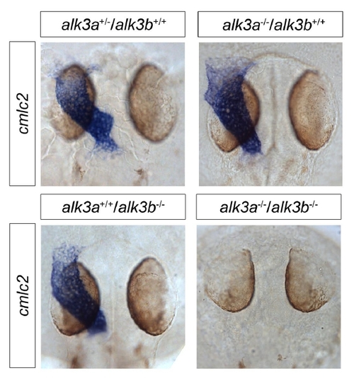
bmpr1aa/bmpr1ab double mutant embryos lack myocardial tissue. In situ hybridization for myl7 (cmlc2) expressed in the myocardium of wild-type, bmpr1aa mutant or bmpr1ab mutant embryos. Myl7 expression was not detected in bmpr1aa/bmpr1ab double mutant embryos. All embryos shown as dorsal views at 30 hpf. |
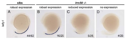
lefty1 expression in lrcc50 mutant embryos. In situ hybridization analysis of lefty1 expression in lrrc50 mutant embryos at 16 somites. The majority of wild type embryos express lefty1 from the posterior tip of the notochord anteriorly to around the middle of the trunk (A). The majority of lrrc50 mutants express lefty1 in a similar domain to wild type embryos (B). A subset of lrrc50 mutants either express lefty1 in a domain restricted to the posterior tip of the notochord (C), or do not expression lefty1 (D). Lateral views, dorsal to the right. |