
- Title
-
HSF1 Is Essential for the Resistance of Zebrafish Eye and Brain Tissues to Hypoxia/Reperfusion Injury
- Authors
- Tucker, N.R., Middleton, R.C., Le, Q.P., and Shelden, E.A.
- Source
- Full text @ PLoS One
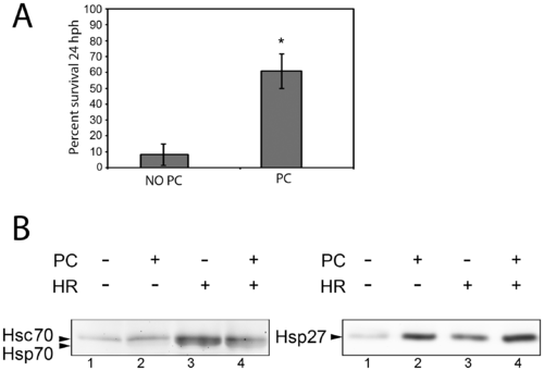
Effect of heat shock preconditioning on embryo survival and heat shock protein expression. A: Average percentage survival of embryos treated at 48 hpf with 160 min of hypoxia. Embryos were either heat shock preconditioned (PC) or not (No PC) and analyzed for survival 24 hours post hypoxia. Data shown are from five independent experiments. Error bars are the SEM. The asterisk indicates a statistically significant difference. B: Representative Western blots showing Hsp70 and Hsp27 expression after heat shock preconditioning and/or hypoxia treatments. EXPRESSION / LABELING:
|

Effects of heat shock preconditioning on cell death in zebrafish brain and eye tissues after sublethal hypoxia treatments. A: The average number of TUNEL-positive nuclei per unit volume in the eye and brain following 100 min of hypoxia at 48 hpf and 8 hours of recovery in normoxic media. Asterisks indicate statistically significant differences between values obtained from non-preconditioned (No PC) embryos and embryos preconditioned with a heat shock at 34 hpf (PC). Error bars are the SEM. B: TUNEL positive cells in representative images of eye (upper panels) and brain (lower panels) regions after hypoxia. Dashed lines indicate the region in which nuclei were counted. Scale bars are 100 μm. Asterisks mark the dorsal, anterior aspect of the embryos. C: Hsp70 and Hsp27 immunostaining of brain (left panels) and eye (right panels) tissues. Arrows highlight regions showing increased Hsp70 staining intensity of heat shock preconditioned (PC) tissues compared to non-preconditioned (No-PC) controls. Increases in Hsp27 staining intensity are also evident throughout the preconditioned tissues. Scale bars are 100 μm. EXPRESSION / LABELING:
|
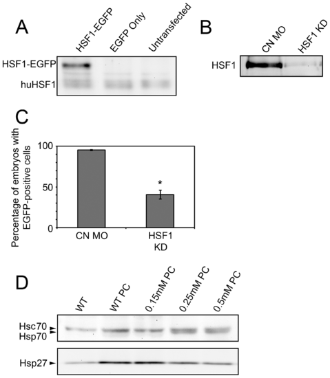
Assessment of HSF1 knockdown following HSF1 MO injection. A: Western blots of HeLa cell protein using the SPA-901 anti-Human HSF1 antibody. A single band is seen at the predicted molecular weight of human HSF1 (huHSF1) in all lanes. An additional higher molecular weight band is detected in extracts of cells transfected with plasmid coding for a zebrafish HSF1-EGFP fusion protein (left lane), but not EGFP alone (middle lane). B: Western blot of HSF1 in nuclear extracts of 48 hpf zebrafish embryos. Embryos were injected with control (CN MO) or anti-HSF1 (HSF1 KD) MO. C: Quantification of the number of embryos containing fluorescent cells following co-injection of a HSF1-EGFP reporter plasmid and control (CN MO) or HSF1 antisense (HSF1 KD) MO. Error bars are the SEM. The asterisk indicates a statistically significant difference between α-HSF1 and CN-MO injected embryos. D: Effect of α-HSF1 MO on heat shock protein expression after heat shock preconditioning (PC). Embryos were injected with the indicated concentrations of α-HSF1 MO or uninjected (WT), followed by Western blotting for the detection of Hsp70 and Hsp27. EXPRESSION / LABELING:
|
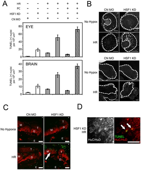
Effects of HSF1 knockdown on apoptosis. A: Quantification of TUNEL positive nuclei in the eye and brain of 58 hpf embryos, showing effects of HSF1 knockdown (compare HSF1 KD versus CN MO), in embryos incubated under control conditions (white bars), after hypoxia/reperfusion only (gray bars), and after heat shock preconditioning followed by hypoxia/reperfusion (striped bars). Error bars are the SEM. B: TUNEL positive cells in representative images of eye (upper panels) and brain (lower panels) regions after hypoxia. Dashed lines indicate the region in which nuclei were counted. Scale bars are 50 μm. C: Analysis of brain morphology and apoptosis after HSF1 knockdown. Representative images showing HuC/HuD immunostaining of neurons (red) and TUNEL positive nuclei (green) in brain sections obtained from embryos injected with control (CN) or HSF1 antisense (HSF1 KD) MO and incubated in normoxic medium (No Hypoxia) or exposed to hypoxia and reperfusion (HR). Asterisks mark the dorsal, anterior aspect of the embryos. Labels are telencephalon (T), midbrain (M), diencephalon (D), and eye (E). Scale bars are 50 μm. D: Representative higher magnification images showing localization of markers for neurons and apoptotic cells in cryosectioned brain. Scale bars are 10 μm. EXPRESSION / LABELING:
|
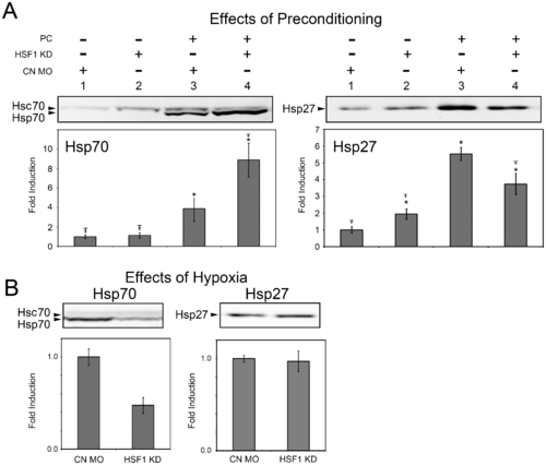
Alterations in heat shock protein expression following HSF1 knockdown. A: Representative Western blots, and quantitative analysis of Hsp70 and Hsp27 expression. Expression was analyzed in 48 hpf embryos injected with control (CN MO) or HSF1 antisense (HSF1 KD) morpholinos incubated at control temperatures or subjected to heat shock preconditioning (PC). Values represent average data from four independent experiments. Error bars are the SD, symbols indicate a significant difference from CN-MO injected embryos that were not preconditioned (*) and from CN-MO injected embryos that were preconditioned (Ŧ–). B: Representative Western blots and quantitative analysis of Hsp27 and Hsp70 expression after a 100 min hypoxia treatment. Embryos were injected with HSF1 antisense MO (HSF1 KD) or a control MO (CN). Bars represent data collected from three independent experiments. Error bars are the SEM. EXPRESSION / LABELING:
|