
Fig. 4
- ID
- ZDB-FIG-120613-6
- Publication
- Tucker et al., 2011 - HSF1 Is Essential for the Resistance of Zebrafish Eye and Brain Tissues to Hypoxia/Reperfusion Injury
- Other Figures
- All Figure Page
- Back to All Figure Page
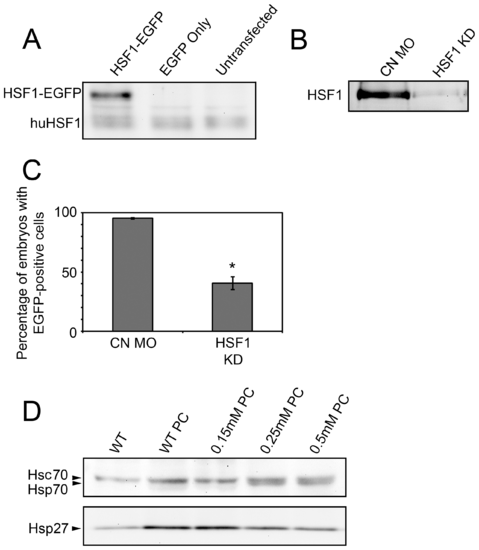
Assessment of HSF1 knockdown following HSF1 MO injection. A: Western blots of HeLa cell protein using the SPA-901 anti-Human HSF1 antibody. A single band is seen at the predicted molecular weight of human HSF1 (huHSF1) in all lanes. An additional higher molecular weight band is detected in extracts of cells transfected with plasmid coding for a zebrafish HSF1-EGFP fusion protein (left lane), but not EGFP alone (middle lane). B: Western blot of HSF1 in nuclear extracts of 48 hpf zebrafish embryos. Embryos were injected with control (CN MO) or anti-HSF1 (HSF1 KD) MO. C: Quantification of the number of embryos containing fluorescent cells following co-injection of a HSF1-EGFP reporter plasmid and control (CN MO) or HSF1 antisense (HSF1 KD) MO. Error bars are the SEM. The asterisk indicates a statistically significant difference between α-HSF1 and CN-MO injected embryos. D: Effect of α-HSF1 MO on heat shock protein expression after heat shock preconditioning (PC). Embryos were injected with the indicated concentrations of α-HSF1 MO or uninjected (WT), followed by Western blotting for the detection of Hsp70 and Hsp27. |
| Antibodies: | |
|---|---|
| Fish: | |
| Condition: | |
| Knockdown Reagent: | |
| Anatomical Term: | |
| Stage: | Long-pec |