
- Title
-
Dependence of cardiac trabeculation on neuregulin signaling and blood flow in zebrafish
- Authors
- Peshkovsky, C., Totong, R., and Yelon, D.
- Source
- Full text @ Dev. Dyn.
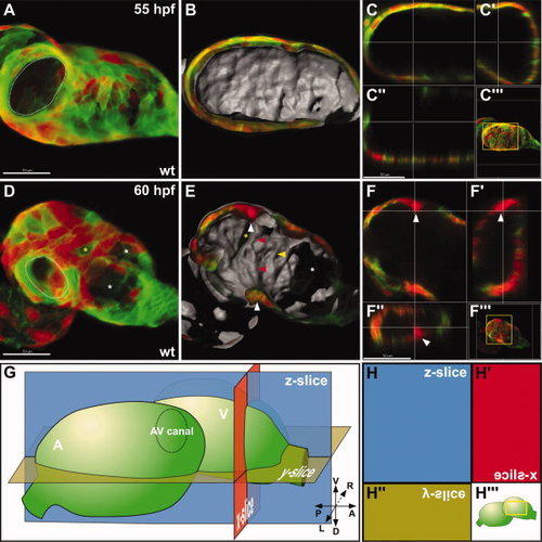
Trabeculation initiates between 55 and 60 hours postfertilization (hpf) in a characteristic location. A–F: Confocal reconstructions of the ventricular myocardium in wild-type (wt) embryos expressing Tg(myl7:dsredt4) and Tg(myl7:egfp-hshras). Volume reconstruction (A,D), lumenal surface reconstruction (B,E), and optical sections (C,F) are shown as described in the Experimental Procedures section. In A and D, the atrioventricular (AV) canal is outlined with a dotted white line. White asterisks indicate areas where the acquisition of fluorescent signal was blocked by overlying melanocytes. Yellow asterisks indicate examples of cells displaying epigenetic silencing of Tg(myl7:dsredt4). G,H: Diagrams depict the orientations of the hearts and optical sections analyzed, as described in the Experimental Procedures section. The axes indicate the orientation of the embryo: V, ventral; D, dorsal; P, posterior; A, anterior; L, left; R, right. Additional abbreviations used are A for atrium and V for ventricle. A–C: At 55 hpf, the lumenal surface of the ventricle is generally smooth with no prominent protrusions. See also Supp. Movie S1. D–F: By 60 hpf, lumenal protrusion becomes evident (E, red arrowheads), most notably at a characteristic position on the ventricular outer curvature (E, top white arrowhead, and F, white arrowheads). Additional portions of the lumenal surface of the chamber exhibit more subtle and variable protrusions (E, yellow arrowhead). See also Supp. Movie S2. Scale bars = 50 μm. |
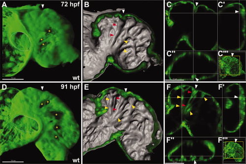
Lumenal ridges become prominent by 3 days postfertilization (dpf). A–F: Confocal reconstructions of the ventricular myocardium in wild-type (wt) embryos expressing Tg(myl7:egfp). All reconstructions and sections are as shown in Figure 1. Yellow asterisks indicate examples of cells displaying epigenetic silencing of Tg(myl7:egfp). White arrowheads indicate the position of the invagination in the ventricular wall. A–C: By 72 hours postfertilization (hpf), ridges are present on a broad portion of the lumenal surface of the ventricle (red and yellow arrowheads). The most prominent ridge (red arrowheads) extends from the atrioventricular (AV) canal to the outer curvature and appears tethered to an invagination in the ventricular wall (white arrowhead). See also Supp. Movie S3. D–F: By 91 hpf, the ridges (red and yellow arrowheads) become more complex, although most ridges form a radial array that coalesce at a common origin near the AV canal. See also Supp. Movie S4. |

The main trabecular stalk separates from the ventricular wall between 96 hours postfertilization (hpf) and 120 hpf. A–F: Confocal reconstructions of the ventricular myocardium in wild-type (wt) embryos expressing Tg(myl7:dsredt4) and Tg(myl7:egfp-hshras) (A–C) or expressing only Tg(myl7:dsredt4) (D–F). All reconstructions and sections are as shown in Figure 1. A–C: By 96 hpf, the main ridge (red arrowheads) on the lumenal surface becomes a thick stalk that is connected to numerous other ridges crossing the chamber (yellow arrowheads). See also Supp. Movie S5. D–F: By 120 hpf, the main stalk (red arrowheads) separates from the ventricular wall, but is still connected to the wall by thin connecting rods (green arrowheads). Additionally, the interconnections between the other ridges (yellow arrowheads) become increasingly more elaborate. See also Supp. Movie S6. |
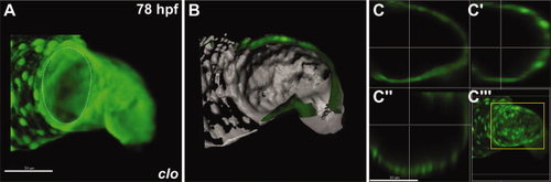
In clo mutant embryos, trabeculation fails to initiate. A–C: Confocal reconstructions of the ventricular myocardium in clo mutant embryos expressing Tg(myl7:egfp). All reconstructions and sections are as shown in Figure 1. A similarly staged wild-type (wt) embryo is shown in Figure 2A–C. A: The clo mutant ventricle is dysmorphic and has a large AV canal. B,C: The lumenal surface of the clo mutant ventricle is smooth with no significant protrusions or ridges, and the ventricular wall retains a simple, uniform thickness. See also Supp. Movie S7. |

Neuregulin signaling is required for the initiation of trabeculation. A–L: Confocal reconstructions of the ventricular myocardium in wild-type (wt) embryos expressing Tg(myl7:dsredt4) and Tg(myl7:egfp-hshras) and treated either with dimethyl sulfoxide (DMSO; A–C,G–I) or AG1478 (D–F,J–L). All reconstructions and sections are as shown in Figure 1, except that the atrioventricular (AV) canal is not visible in G and J. A white asterisk indicates area where the acquisition of fluorescent signal was blocked by overlying melanocytes. Yellow asterisks indicate examples of cells displaying epigenetic silencing of Tg(myl7:dsredt4). A–C: At 75 hours postfertilization (hpf), DMSO-treated embryos exhibit lumenal protrusions and primitive ridges (yellow arrowheads), similar to untreated wt embryos at a comparable stage (Fig. 2A–C). See also Supp. Movie S8. D–F: In contrast, AG1478-treated embryos do not initiate trabeculation and instead exhibit a smooth lumenal surface and a uniform thickness of the chamber wall at 75 hpf. See also Supp. Movie S9. G–I: At 99 hpf, DMSO-treated embryos exhibit normal ventricular morphology, including defined trabecular ridges (yellow arrowheads). See also Supp. Movie S10. J–L: In contrast, AG1478-treated embryos fail to form defined myocardial protrusions or ridges, although some subtle and variable displacements of lumenal cardiomyocytes do emerge (K, yellow arrowheads). See also Supp. Movie S11. |
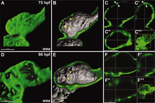
Reduced blood flow in wea mutant embryos inhibits the progression of trabeculation. A–F: Confocal reconstructions of the ventricular myocardium in wea mutant embryos expressing Tg(myl7:egfp). Using a stereomicroscope, we confirmed the presence of suboptimal blood flow through the ventricle in all wea mutant embryos chosen for analysis. All reconstructions and sections are as shown in Figure 1. Similarly staged wild-type (wt) embryos are shown in Figure 2. White asterisks indicate areas where the acquisition of fluorescent signal was blocked by overlying melanocytes. Yellow asterisks indicate examples of cells displaying epigenetic silencing of Tg(myl7:egfp). A: At 75 hours postfertilization (hpf), the wea mutant ventricle is dysmorphic and has a narrow AV canal. B,C: A few cells on the lumenal surface of the wea mutant ventricle appear to protrude into the chamber (B, yellow arrowheads) and the ventricular wall exhibits some areas of subtle thickening (C, white arrowheads). See also Supp. Movie S12. D–F: At 96 hpf, the wea mutant ventricle lacks ridges or trabeculae; however, some protruding cells are still present (E, yellow arrowheads). See also Supp. Movie S13. |