
|
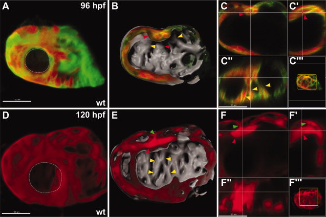
Fig. 3 The main trabecular stalk separates from the ventricular wall between 96 hours postfertilization (hpf) and 120 hpf. A–F: Confocal reconstructions of the ventricular myocardium in wild-type (wt) embryos expressing Tg(myl7:dsredt4) and Tg(myl7:egfp-hshras) (A–C) or expressing only Tg(myl7:dsredt4) (D–F). All reconstructions and sections are as shown in Figure 1. A–C: By 96 hpf, the main ridge (red arrowheads) on the lumenal surface becomes a thick stalk that is connected to numerous other ridges crossing the chamber (yellow arrowheads). See also Supp. Movie S5. D–F: By 120 hpf, the main stalk (red arrowheads) separates from the ventricular wall, but is still connected to the wall by thin connecting rods (green arrowheads). Additionally, the interconnections between the other ridges (yellow arrowheads) become increasingly more elaborate. See also Supp. Movie S6.