
Fig. 2
- ID
- ZDB-FIG-110216-18
- Publication
- Peshkovsky et al., 2011 - Dependence of cardiac trabeculation on neuregulin signaling and blood flow in zebrafish
- Other Figures
- All Figure Page
- Back to All Figure Page
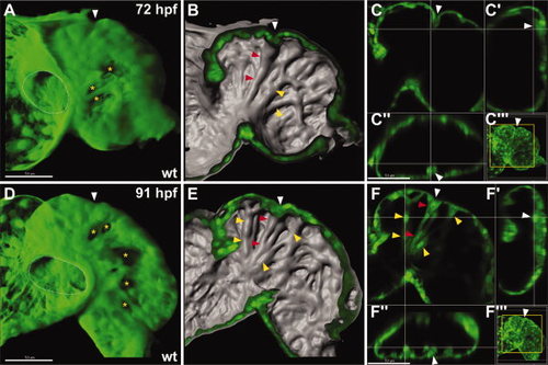
Lumenal ridges become prominent by 3 days postfertilization (dpf). A–F: Confocal reconstructions of the ventricular myocardium in wild-type (wt) embryos expressing Tg(myl7:egfp). All reconstructions and sections are as shown in Figure 1. Yellow asterisks indicate examples of cells displaying epigenetic silencing of Tg(myl7:egfp). White arrowheads indicate the position of the invagination in the ventricular wall. A–C: By 72 hours postfertilization (hpf), ridges are present on a broad portion of the lumenal surface of the ventricle (red and yellow arrowheads). The most prominent ridge (red arrowheads) extends from the atrioventricular (AV) canal to the outer curvature and appears tethered to an invagination in the ventricular wall (white arrowhead). See also Supp. Movie S3. D–F: By 91 hpf, the ridges (red and yellow arrowheads) become more complex, although most ridges form a radial array that coalesce at a common origin near the AV canal. See also Supp. Movie S4. |