
|
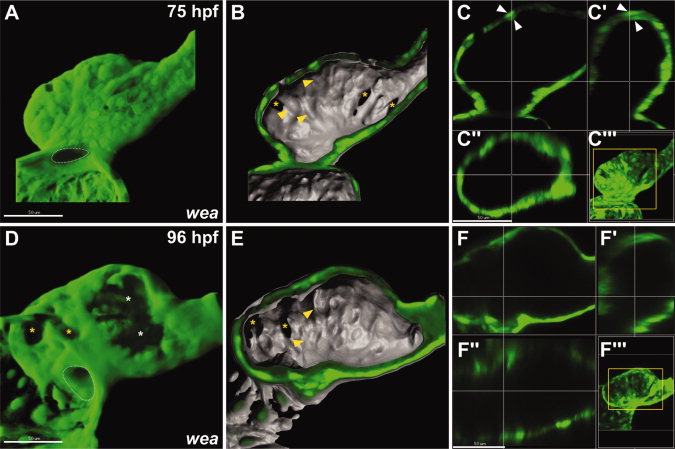
Fig. 6 Reduced blood flow in wea mutant embryos inhibits the progression of trabeculation. A–F: Confocal reconstructions of the ventricular myocardium in wea mutant embryos expressing Tg(myl7:egfp). Using a stereomicroscope, we confirmed the presence of suboptimal blood flow through the ventricle in all wea mutant embryos chosen for analysis. All reconstructions and sections are as shown in Figure 1. Similarly staged wild-type (wt) embryos are shown in Figure 2. White asterisks indicate areas where the acquisition of fluorescent signal was blocked by overlying melanocytes. Yellow asterisks indicate examples of cells displaying epigenetic silencing of Tg(myl7:egfp). A: At 75 hours postfertilization (hpf), the wea mutant ventricle is dysmorphic and has a narrow AV canal. B,C: A few cells on the lumenal surface of the wea mutant ventricle appear to protrude into the chamber (B, yellow arrowheads) and the ventricular wall exhibits some areas of subtle thickening (C, white arrowheads). See also Supp. Movie S12. D–F: At 96 hpf, the wea mutant ventricle lacks ridges or trabeculae; however, some protruding cells are still present (E, yellow arrowheads). See also Supp. Movie S13.