
- Title
-
Distinct troponin C isoform requirements in cardiac and skeletal muscle
- Authors
- Sogah, V.M., Serluca, F.C., Fishman, M.C., Yelon, D.L., MacRae, C.A., and Mably, J.D.
- Source
- Full text @ Dev. Dyn.
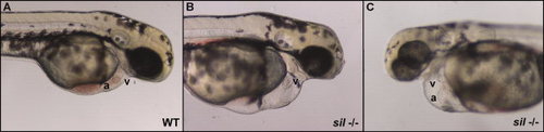
Characterization of the sil phenotype. Lateral view of wild-type (WT) (A) and silm656 mutant (-/-) (B, C) embryos at 2 dpf. The ventricle (v) of the sil mutant embryo (B, C) is visibly compacted and misshapen compared to wild-type (A), while the atrium (a) of the sil mutant (C) has become enlarged. The silm656 mutant embryo (B, C) also exhibits pericardial edema and has reduced circulation as compared to wild-type (A). Mutant embryos homozygous for the silsk25 allele exhibit the same phenotype. PHENOTYPE:
|
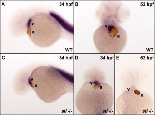
The sil mutant exhibits a loss of expression of ventricular contractile protein genes. Double staining with an atrial-specific antibody (S46) and an in situ RNA probe for ventricular myosin heavy chain (vmhc) (Yelon et al., 1999) was used to distinguish the atrial and ventricular chambers in wild-type (WT) (A, B) and silm656 mutant (-/-) (C-E) embryos. The hearts of wild-type (A) and silm656 mutant (C, D) embryos at 34 hpf express markers specific for the two cardiac chambers. However, by 52 hpf, the hearts of silm656 mutant (E) embryos no longer express the ventricle-specific vmhc marker while both chamber markers are still visible in wild-type embryos (B). |
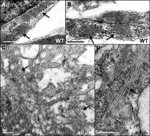
Myofibrillar structure is disorganized in the sil mutant. Transmission electron microscopy was performed to study the structure of ventricular cardiomyocytes from wild-type (WT) (A, B) and silm656 mutant (-/-) (C, D) embryos at 50 hpf. A, B: Myofibrils are easily detectable in the cardiomyocytes from ventricles of wild-type embryos. C, D: silm656 mutant embryos exhibit very sparse and poorly organized sarcomeres. Arrows are used to indicate myofibrils. Bars = 500 nm. PHENOTYPE:
|

Expression of tnnc1a, tnnc1b, and tnnc2 mRNA in zebrafish. A, B: The zebrafish tnnc1a gene is expressed throughout the heart of wild-type zebrafish embryos at 30 hpf, indicated by arrows. C: By 50 hpf, tnnc1a expression is predominantly restricted to the ventricle (v) of wild-type hearts and excluded from the atrium (a). The tnnc1a gene is expressed in cardiac tissue (indicated by the arrow) at both 30 hpf (D) and 50 hpf (D′). The second cTnC gene in zebrafish (tnnc1b) is detected predominantly in skeletal muscle (indicated by arrows) at 30 hpf (E) with decreased expression by 50 hpf (E′). The sTnC gene in zebrafish (tnnc2) is expressed in skeletal muscle (indicated by arrows) at both 30 hpf (F) and 50 hpf (F′). |
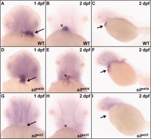
In situ analysis of tnnc1a mRNA expression in sil mutant embryos. In silm656 mutant embryos at 1 dpf (D), tnnc1a expression is found throughout the heart as in wild-type (WT) embryos (A). However, by 2 dpf, the expression of tnnc1a in silm656 embryos (E, F) has become diffuse compared to wild-type (B, C). No tnnc1a expression can be detected in silsk25 mutant embryos either at 1 or 2 dpf (G-I). |
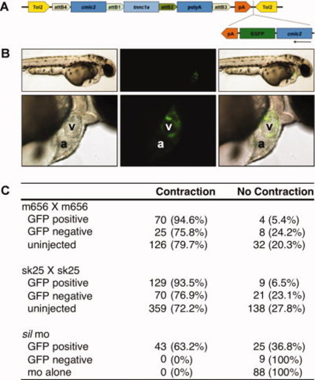
Myocardial-specific tnnc1a expression rescues the sil mutant phenotype. A: Depiction of the tnnc1a rescue construct used for injection of zebrafish embryos at the 1-2-cell stage. B: 2-dpf wild-type embryos coinjected with tnnc1a morpholino (MO), the rescue construct depicted in A, and transposase RNA. The top row shows the whole embryo and the bottom row shows the heart of the same embryo magnified 5×. GFP indicates expression of the construct in the cardiomyocytes of this embryo. C: Table summarizing the results of rescue experiments in the silm656 line, the silsk25 line, and wild-type embryos injected with tnnc1a MO. Uninjected controls are compared with injected embryos that either have at least one GFP-positive cell (GFP positive) or no GFP-positive cells (GFP negative). |
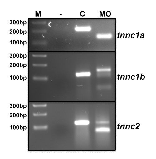
Confirmation of altered splice site use in tnnc1a, tnnc1b, and tnnc2 morpholino-injected embryos. mRNA was isolated from pooled embryos at 1 dpf and analyzed using primers flanking the morpholino-targeted exon donor site. PCR was performed using the Qiagen OneStep RT-PCR kit. Lane 1: No DNA control; Lane 2: uninjected embryos; Lane 3: morpholino-injected embryos. Embryos were injected with 200 μM tnnc1a, 200 μM tnnc1b, or 500 μM tnnc2 morpholino. |
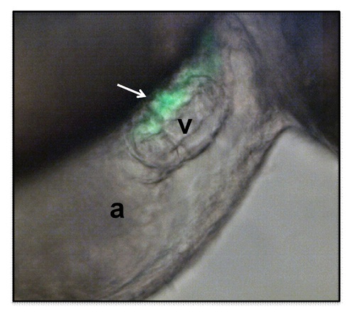
Rescue of the silsk25 mutant phenotype by myocardial specific tnnc1a expression. Overlay of DIC and FITC channel of a frame from Supp. Movie S3 demonstrating expression of the cmlc2::tnnc1a/cmlc2::GFP transgene on the dorsal surface of the ventricle. The GFP expression correlates with the region of rescued myocardium. |