
Image
|
Figure Caption
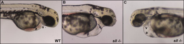
Fig. 1 Characterization of the sil phenotype. Lateral view of wild-type (WT) (A) and silm656 mutant (-/-) (B, C) embryos at 2 dpf. The ventricle (v) of the sil mutant embryo (B, C) is visibly compacted and misshapen compared to wild-type (A), while the atrium (a) of the sil mutant (C) has become enlarged. The silm656 mutant embryo (B, C) also exhibits pericardial edema and has reduced circulation as compared to wild-type (A). Mutant embryos homozygous for the silsk25 allele exhibit the same phenotype.
Figure Data
Acknowledgments
This image is the copyrighted work of the attributed author or publisher, and
ZFIN has permission only to display this image to its users.
Additional permissions should be obtained from the applicable author or publisher of the image.
Full text @ Dev. Dyn.