
Image
|
Figure Caption
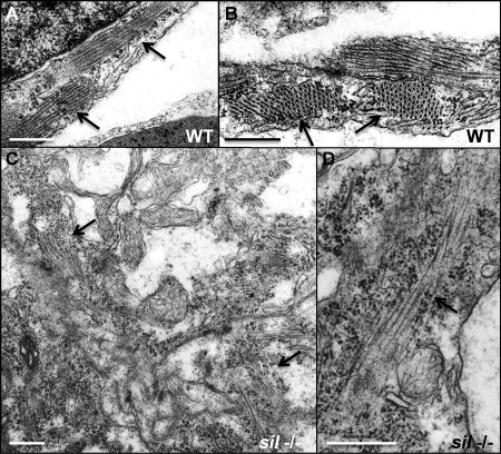
Fig. 3 Myofibrillar structure is disorganized in the sil mutant. Transmission electron microscopy was performed to study the structure of ventricular cardiomyocytes from wild-type (WT) (A, B) and silm656 mutant (-/-) (C, D) embryos at 50 hpf. A, B: Myofibrils are easily detectable in the cardiomyocytes from ventricles of wild-type embryos. C, D: silm656 mutant embryos exhibit very sparse and poorly organized sarcomeres. Arrows are used to indicate myofibrils. Bars = 500 nm.
Figure Data
Acknowledgments
This image is the copyrighted work of the attributed author or publisher, and
ZFIN has permission only to display this image to its users.
Additional permissions should be obtained from the applicable author or publisher of the image.
Full text @ Dev. Dyn.