
FIGURE
Fig. 3
- ID
- ZDB-FIG-101105-21
- Publication
- Sogah et al., 2010 - Distinct troponin C isoform requirements in cardiac and skeletal muscle
- Other Figures
- All Figure Page
- Back to All Figure Page
Fig. 3
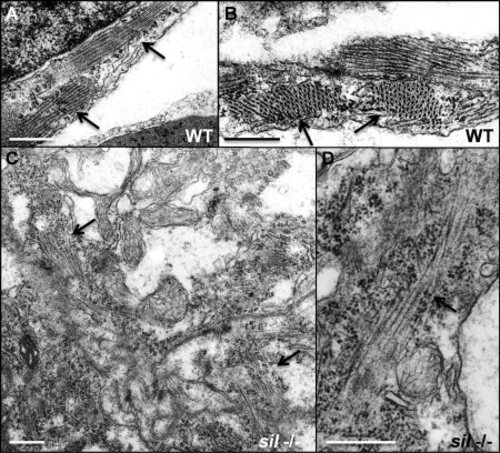
Myofibrillar structure is disorganized in the sil mutant. Transmission electron microscopy was performed to study the structure of ventricular cardiomyocytes from wild-type (WT) (A, B) and silm656 mutant (-/-) (C, D) embryos at 50 hpf. A, B: Myofibrils are easily detectable in the cardiomyocytes from ventricles of wild-type embryos. C, D: silm656 mutant embryos exhibit very sparse and poorly organized sarcomeres. Arrows are used to indicate myofibrils. Bars = 500 nm. |
Expression Data
Expression Detail
Antibody Labeling
Phenotype Data
| Fish: | |
|---|---|
| Observed In: | |
| Stage: | Long-pec |
Phenotype Detail
Acknowledgments
This image is the copyrighted work of the attributed author or publisher, and
ZFIN has permission only to display this image to its users.
Additional permissions should be obtained from the applicable author or publisher of the image.
Full text @ Dev. Dyn.