
- Title
-
Fish-specific duplicated dmrt2b contributes to a divergent function through Hedgehog pathway and maintains left-right asymmetry establishment function
- Authors
- Liu, S., Li, Z., and Gui, J.F.
- Source
- Full text @ PLoS One

Molecular characterization and expression pattern of zebrafish Dmrt2b during embryogenesis. (A) Amino acid alignment of zebrafish Dmrt2b with gibel carp Dmrt2b, gibel carp Dmrt2a and zebrafish Dmrt2a. Similar and identical amino acids are highlighted in grey and black boxes. The line indicates the DM domain. (B) RT-PCR detection of dmrt2b in zebrafish embryonic development stages, and beta-actin mRNA as the control. (C) Western blot detection of Dmrt2b during the zebrafish embryo development, and Tubulin was used for the control. (D-H) Whole-mount in situ hybridization detection of dmrt2b on somitogenesis embryos as indicated stage. The arrows indicate positive signals in the somites. |
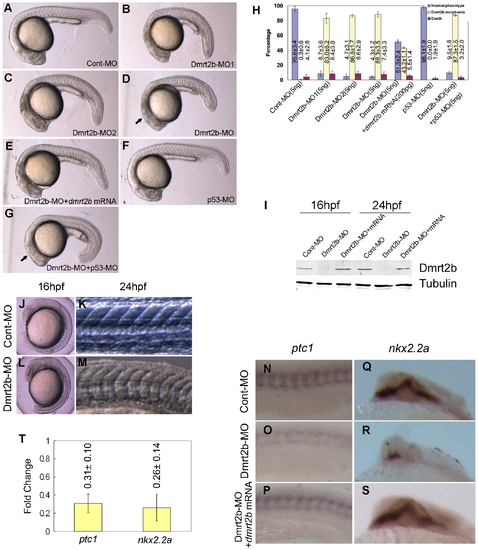
Dmrt2b morphants display defects in somitogenesis and Hedgehog signaling. (A–G) Morphology of 24 hpf embryos injected with Cont-MO (A), Dmrt2b-MO1 (B), Dmrt2b-MO2 (C), Dmrt2b-MO (D), Dmrt2b-MO+dmrt2b mRNA (E), p53-MO (F), Dmrt2b-MO+p53-MO (G). The arrows indicate the off-target cell death in Dmrt2b-MO morphant and the reduced cell death in the Dmrt2b-MO+p53-MO morphant. (H) The statistical data of three independent experiments on Dmrt2b knockdown, dmrt2b mRNA rescue and p53 MO co-injection. Results are represented as mean±SD of three separate experiments. (I) Western blot detection of Dmrt2b knockdown during embryogenesis. The protein extracts from embryos (16 hpf and 24 hpf) were analyzed by Western blot using the polyclonal anti-Dmrt2b antibody. A band of about 41 KD was not detected in Dmrt2b morphants. The picture represents typical result from three separate experiments. (J–M) Dmrt2b morphant exhibits U-shape somites. Morphology of embryos injected with Cont-MO display the typical ‘chevron’ shape (J, K). Morphology of embryos injected with Dmrt2b-MO display the U-shape (L, M). Whole-mount in situ hybridization of ptc1(N, O, P) (Anterior is left) and nkx2.2a(Q, R, S) (Anterior is top) in embryos injected with Cont-MO (N, Q) or Dmrt2b-MO (O, R) and embryos co-injected with Dmrt2b-MO with dmrt2b mRNA (P, S) at 24 hpf. (T) qPCR analysis of the expression changes of ptc1 and nkx2.2a in 24 hpf embryos injected with Cont-MO or Dmrt2b-MO. Results represent mean±SD of three separate experiments. |
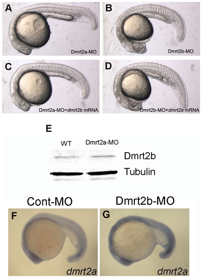
Dmrt2a and Dmrt2b can not compensate for each other. (A–D)Morphology of 24 hpf embryos injected with Dmrt2a-MO (A), Dmrt2b-MO (B), Dmrt2a-MO+dmrt2b mRNA (C), Dmrt2b-MO+dmrt2a mRNA (D). (E) Western blot assay showing Dmrt2b expression level in the embryos injected with wild type (WT) and Dmrt2a-MO. (F, G) The expression of dmrt2a(terra) was not affected in Dmrt2b morphants. Lateral views of embryos at 18 somites stage. EXPRESSION / LABELING:
|
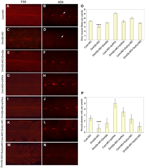
Dmrt2b is involved in slow muscle development through Hedgehog signaling. Analysis of slow muscle development by whole-mount immunostaining of 26 hpf embryos with antibodies labeling slow MyHC (F59) (A, C, E, G, I, K, M) and engrailed muscle pioneer cells (4d9) (B, D, F, H, J, L, N). Lateral view of embryos injected with Cont-MO (A, B), Dmrt2b-MO (C, D), embryos co-injected with Dmrt2b-MO+dmrt2b (E, F), Cont-MO+dnPKA (G, H), Dmrt2b-MO+dnPKA (I, J), Cont-MO+Su(fu)-MO (K, L), Dmrt2b-MO+Su(fu)-MO (M, N). White arrows in 4d9 panel indicate reduction of engrailed staining in the knockdown embryos compared to controls. All images show the somites over the yolk extension. Anterior is left in all images. (O) Quantitative analysis of slow muscle fiber number per somite in embryos at 26 hpf, injected as indicated. Data represent average±SD. *** indicates significance of p<0.0001 for Dmrt2b-MO vs. Cont-MO, Dmrt2b-MO+dmrt2b, Dmrt2b-MO+dnPKA and Dmrt2b-MO+Su(fu)-MO. n = 20 to 38 embryos per condition. (P) Quantitative analysis of muscle pioneer cells number per somite in embryos at 26 hpf, injected as indicated. Data represent average±SD. *** indicates significance of p<0.0001 for Dmrt2b-MO vs. Cont-MO, Dmrt2b-MO+dmrt2b and Dmrt2b-MO+dnPKA. n = 20 to 36 embryos per condition. |
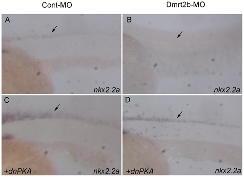
Patterning defects in the neural tube of Dmrt2b morphant embryos. Lateral views of embryos at 24 hpf, anterior to the left, dorsal to the top. (A) Wild phenotype of Cont-MO injected embryo; (B) Severe suppression of nkx2.2a in the neural tube of Dmrt2b-MO injected embryo; (C) Ectopic expression of nkx2.2a in the neural tube of dnPKA injected embryo; (D) The normalized expression of nkx2.2a in the neural tube of dnPKA and Dmrt2b-MO coinjected embryo. Black arrows indicate the expression position of nkx2.2a in the neural tube. All images show the somites over the yolk extension. EXPRESSION / LABELING:
|
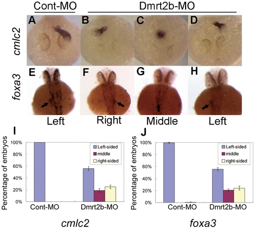
Dmrt2b morphant embryos display defects in heart and visceral organs asymmetry. (A–D) Expression of cmlc2 in 48 hpf embryos detected by whole mount in situ hybridization with antisense RNA probe. (E–H) Expression of foxa3 in 48 hpf embryos detected by whole mount in situ hybridization. Arrows in foxa3 panel indicate the liver. Graphical representation of the percentage of embryos exhibiting the expression patterns for cmlc2 (I) and for foxa3 (J). Results are represented as mean±SD of three separate experiments. |

Dmrt2b knockdown disrupts L–R identity in lateral plate mesoderm. (A–D) lefty1 normal expression in left dorsal diencephalon at 22 somites stage were disrupted embryos injected with Dmrt2b-MO. (E–H) lefty1 normal expression in left LPM at 22 somites stage were disrupted embryos injected with Dmrt2b-MO. (Q) Graphical representation of the percentage of embryos exhibiting the expression patterns for lefty1 in Dmrt2b knockdown embryos and control embryos. (I–L) spaw normal expression in left LPM at 20 somites stage were disrupted embryos injected with Dmrt2b-MO. (R) Graphical representation of the percentage of embryos exhibiting the expression patterns for spaw in Dmrt2b knockdown embryos and control embryos. (M–P) pitx2c normal expression in left LPM at 22 somites stage were disrupted embryos injected with Dmrt2b-MO. (S) Graphical representation of the percentage of embryos exhibiting the expression patterns for pitx2c in Dmrt2b knockdown embryos and control embryos. (T) Lateral view of the embryos at 22 somites stage. Expression of the ntl in embryos injected with Dmrt2b-MO is similar to expression of ntl in control embryos. Results are represented as mean±SD of three separate experiments. (U) qPCR analysis of the expression changes of ntl in 24 hpf embryos injected with Cont-MO or Dmrt2b-MO. The data represents mean±SD of three separate experiments. EXPRESSION / LABELING:
|
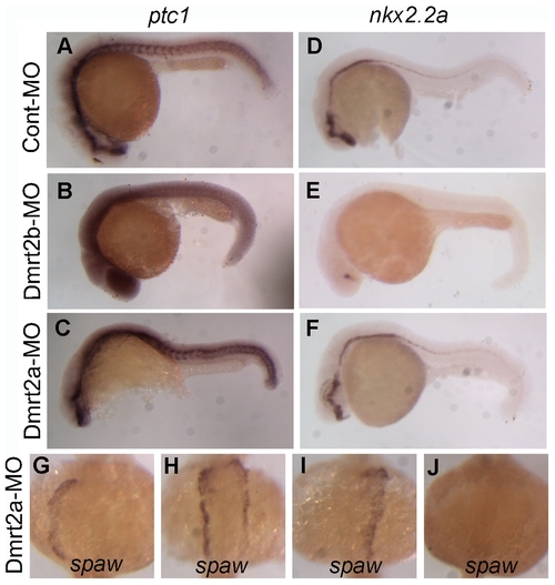
Dmrt2a does not contribute to Hedgehog pathway during zebrafish somitogenesis. (A–C) The expression of ptc1 in embryos injected with Cont-MO, Dmrt2b-MO and Dmrt2a-MO at 24 hpf. (D–F) The expression of nkx2.2a in embryos injected with Cont-MO, Dmrt2b-MO and Dmrt2a-MO at 24 hpf. (G–J) spaw normal expression in left LPM at 20 somites stage were disrupted embryos injected with Dmrt2b-MO. |
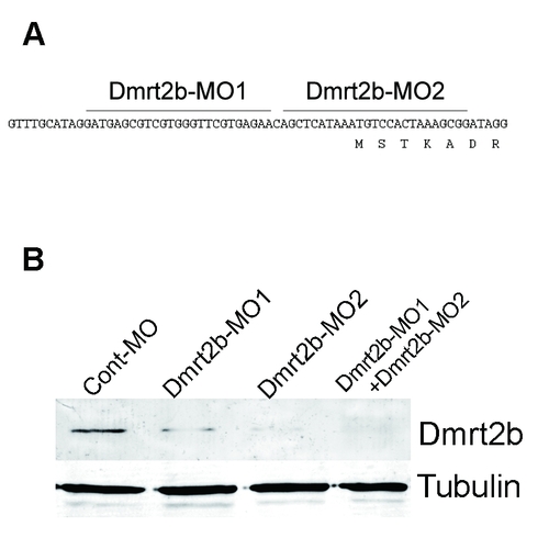
Dmrt2b translation is blocked by non-overlapping morpholinos. (A) The Dmrt2b-MO1 and Dmrt2b-MO2 target sequences are shown in relation to the 5′UTR region of the Dmrt2b mRNA sequence. (B) Western blot assay showing Dmrt2b translation in the embryos injected with Cont-MO, Dmrt2b-MO1, Dmrt2b-MO2 and Dmrt2b-MO (Dmrt2b-MO1+Dmrt2b-MO2). The signal of Dmrt2b protein were significant reduced in Dmrt2b-MO1, Dmrt2b-MO2 and Dmrt2b-MO injected embryos. |

Dmrt2b is not required for shha, ihhb and shhb transcription. Dorsal views of embryos at the bud stage (10 hpf) (A, C, E, G, I and K). Lateral views of embryos at 24 hpf (B, D, F, H, J and L). Expression of shha in embryos injected with Dmrt2b-MO (C and D) is similar to expression of shha in control embryos (A and B). Expression of ihhb in embryos injected with Dmrt2b-MO (G and H) is similar to expression of ihhb in control embryos (E and F). Expression of shhb in embryos injected with Dmrt2b-MO (K and L) is similar to expression of shhb in control embryos (I and J). |
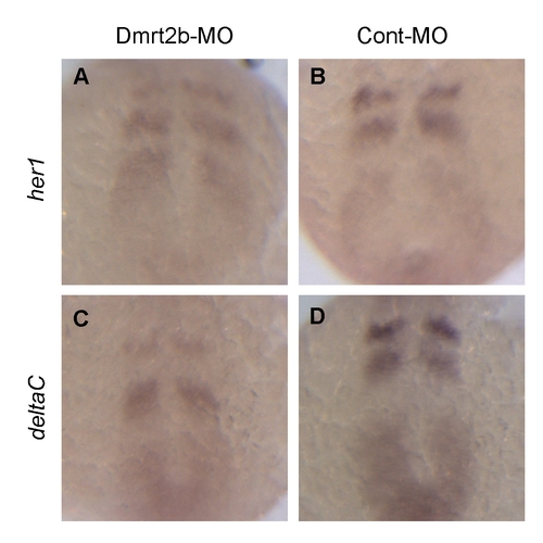
Whole-mount in situ hybridization of persomitic mesoderm genes her1 and deltaC in the Dmrt2b morphants. Expression patterns of her1 in the Dmrt2b-MO (A) and Cont-MO (B) embryos. Expression patterns of deltaC in the Dmrt2b-MO (C) and Cont-MO (D) embryos. All the embryos are at 10 somites stage. Panels show dorsal views, anterior to the top. |