
- Title
-
2,3,7,8-Tetrachlorodibenzo-p-dioxin Exposure Prevents Cardiac Valve Formation in Developing Zebrafish
- Authors
- Mehta, V., Peterson, R.E., and Heideman, W.
- Source
- Full text @ Toxicol. Sci.
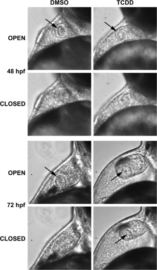
TCDD prevents closure of the nascent AV valve during systole. Newly fertilized zebrafish eggs were treated with TCDD or DMSO as a vehicle control as described in the "Materials and Methods." Representative lateral view micrographs are shown for hearts at the indicated time in development. The arrows indicate an open passage between the atrium and ventricle. Open: the image was taken during diastole in which the AV junction is relaxed. Closed: the image was taken during systole, during which the AV junction has contracted to its fullest extent. An open passage between the ventricle and atrium was consistently observed associated with retrograde blood movement at 72 hpf (see supplemental movie: 72hpfAB.mov). |
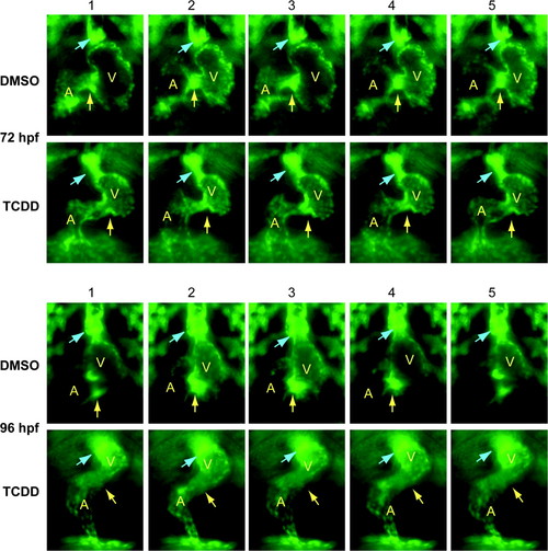
TCDD alters the arrangement of endothelial cell clusters at valve sites. Newly fertilized Tg(flk1:GFP) eggs were treated with TCDD or DMSO as a vehicle control as described in the "Materials and Methods." The figure shows sequential frames taken at six images per second. Representative ventral view fluorescence micrographs showing endocardial cells at the AV and BV junctions are shown for 72 and 96 hpf as indicated. A indicates atrium; V indicates ventricle. Arrows show the location of the AV and BV junctions. The arrows are positioned to indicate forming valve leaflets at the junctions in the DMSO images; leaflets were not observed in the TCDD-exposed hearts. EXPRESSION / LABELING:
|
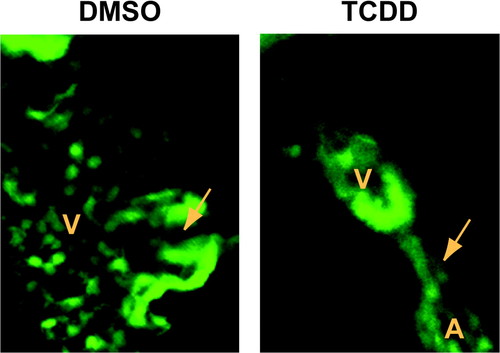
Valve leaflets fail to form in 120 hpf zebrafish exposed to TCDD. Newly fertilized Tg(flk1:GFP) eggs were treated with TCDD or DMSO as a vehicle control and ventral view confocal micrographs were recorded at 120 hpf as described in the Materials and Methods. Representative images show the AV junction, indicated by the arrow. Valve leaflets can be clearly seen flanking the AV passage in the DMSO heart. A indicates atrium; V indicates ventricle. EXPRESSION / LABELING:
|
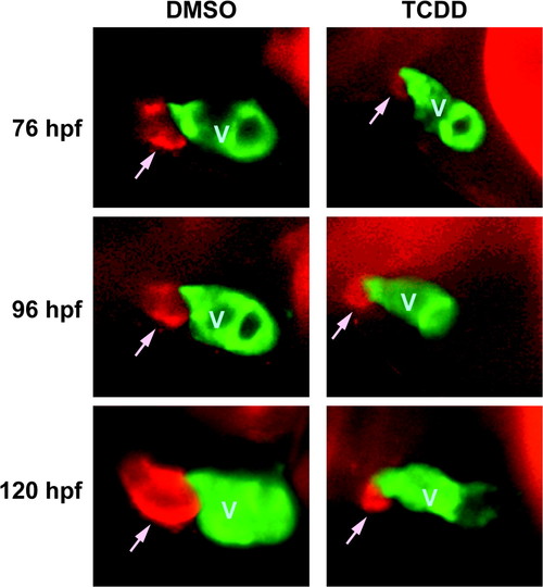
Development of the BA is inhibited by TCDD. Tg(cmlc2::gfp) embryos were exposed to TCDD or DMSO vehicle and treated with DAR-4M AM to stain the BA as described in the Materials and Methods. Embryos were mounted in methylcellulose and visualized using epifluorescence. Lateral view images of representative vehicle and TCDD embryos are shown for 76, 96, and 120 hpf are shown. The head is toward the left of the figure, and the dorsal side is to the top. The ventricle is labeled with GFP, and indicated by a V; the DAR-4 signal identifying the BA is seen as red, and is indicated by an arrow. EXPRESSION / LABELING:
|
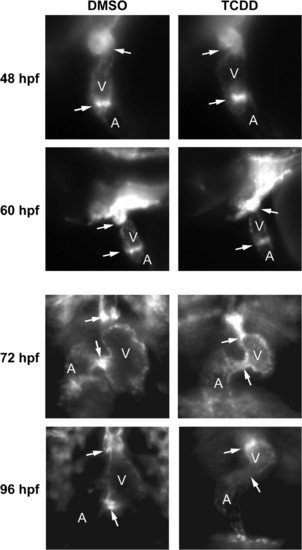
TCDD alters endothelial cell localization at the AV and BV junctions. Newly fertilized Tg(flk1:GFP) eggs were treated with TCDD or DMSO as a vehicle control as described in the "Materials and Methods." Representative fluorescence micrographs showing lateral views (48 and 60 hpf) and ventral views (72 and 96 hpf) of the heart are shown. These orientations were selected to give the best view of the heart at each stage of development. A indicates atrium; V indicates ventricle. Arrows show the locations of the AV and BV junctions. |
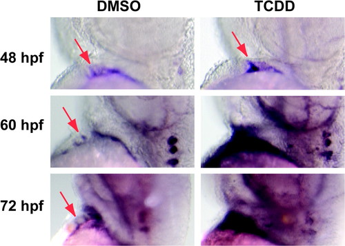
TCDD alters bmp4 mRNA localization in the developing heart. Newly fertilized zebrafish eggs were treated with TCDD or DMSO as a vehicle control as described in the "Materials and Methods." Representative lateral view micrographs are shown for hearts stained by in situ hybridization with a probe specific for bmp4 mRNA. Arrows indicate bmp4 signal at the location of the AV boundary myocardium. The head is toward the top of each panel and the dorsal side is to the right. EXPRESSION / LABELING:
|
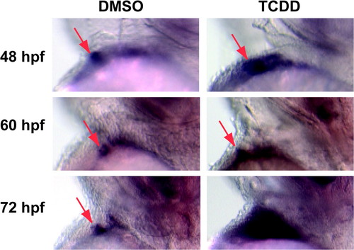
TCDD alters notch1b mRNA localization in the developing heart. Newly fertilized zebrafish eggs were treated with TCDD or DMSO as a vehicle control as described in the "Materials and Methods." Representative lateral view micrographs are shown for hearts stained by in situ hybridization with a probe specific for notch1b mRNA. Arrows indicate notch1b signal at the location of the AV boundary endocardium. The head is toward the top of each panel and the dorsal side is to the right. EXPRESSION / LABELING:
|