
|
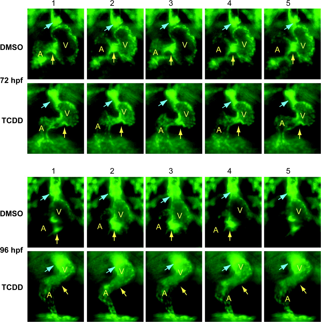
Fig. 3 TCDD alters the arrangement of endothelial cell clusters at valve sites. Newly fertilized Tg(flk1:GFP) eggs were treated with TCDD or DMSO as a vehicle control as described in the "Materials and Methods." The figure shows sequential frames taken at six images per second. Representative ventral view fluorescence micrographs showing endocardial cells at the AV and BV junctions are shown for 72 and 96 hpf as indicated. A indicates atrium; V indicates ventricle. Arrows show the location of the AV and BV junctions. The arrows are positioned to indicate forming valve leaflets at the junctions in the DMSO images; leaflets were not observed in the TCDD-exposed hearts.