
Image
|
Figure Caption
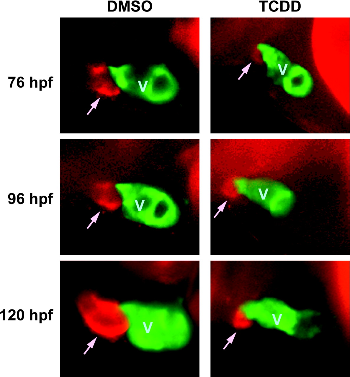
Fig. 5 Development of the BA is inhibited by TCDD. Tg(cmlc2::gfp) embryos were exposed to TCDD or DMSO vehicle and treated with DAR-4M AM to stain the BA as described in the Materials and Methods. Embryos were mounted in methylcellulose and visualized using epifluorescence. Lateral view images of representative vehicle and TCDD embryos are shown for 76, 96, and 120 hpf are shown. The head is toward the left of the figure, and the dorsal side is to the top. The ventricle is labeled with GFP, and indicated by a V; the DAR-4 signal identifying the BA is seen as red, and is indicated by an arrow.
Figure Data
Acknowledgments
This image is the copyrighted work of the attributed author or publisher, and
ZFIN has permission only to display this image to its users.
Additional permissions should be obtained from the applicable author or publisher of the image.
Open access.
Full text @ Toxicol. Sci.