
Image
|
Figure Caption
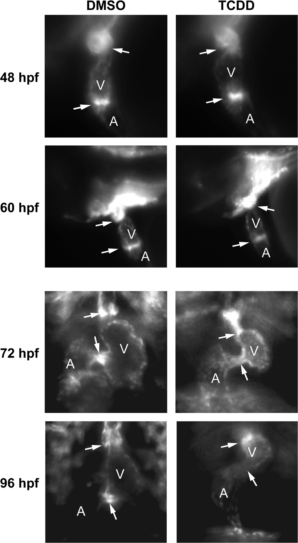
Fig. 6 TCDD alters endothelial cell localization at the AV and BV junctions. Newly fertilized Tg(flk1:GFP) eggs were treated with TCDD or DMSO as a vehicle control as described in the "Materials and Methods." Representative fluorescence micrographs showing lateral views (48 and 60 hpf) and ventral views (72 and 96 hpf) of the heart are shown. These orientations were selected to give the best view of the heart at each stage of development. A indicates atrium; V indicates ventricle. Arrows show the locations of the AV and BV junctions.
Figure Data
Acknowledgments
This image is the copyrighted work of the attributed author or publisher, and
ZFIN has permission only to display this image to its users.
Additional permissions should be obtained from the applicable author or publisher of the image.
Open access.
Full text @ Toxicol. Sci.