
- Title
-
Time-dependent patterning of the mesoderm and endoderm by Nodal signals in zebrafish
- Authors
- Hagos, E.G., and Dougan, S.T.
- Source
- Full text @ BMC Dev. Biol.
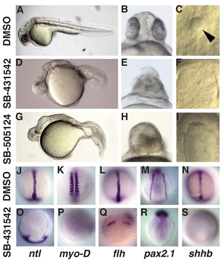
Treatment with 800 μM SB-431542 or 50 μM SB-505124 at MBT prevents formation of mesoderm and endoderm. (A-F) Images of live embryos at 24hpf treated at 2.75 h with DMSO (A-C), SB-431542 (D-F; J-S), or SB-505124 (G-I). Embryos treated with SB-431542 (D-F) or SB-505124 (G-I) lack derivatives of the mesoderm and endoderm in the head and trunk, display severe cyclopia and lack Kupffer's vesicle. (J-P) Images of embryos treated with DMSO (J-N) or SB-431542 at MBT (O-S) and processed to reveal expression of markers for derivatives of dorsal mesoderm (ntl: J, O; flh: L, Q), paraxial mesoderm (myoD: K, P), intermediate mesoderm (pax2.1: M, R), and ventral neurectoderm (shhb: N, S). Dorsal views of embryos fixed at 10 h (J, L, N, P, Q, S)or 14 h (K, M, P, R). Arrowhead in (C) is the Kupffer's vesicle. EXPRESSION / LABELING:
|
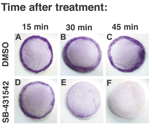
SB-431542 rapidly blocks transcription of Nodal target genes. lefty1 expression in embryos treated with DMSO (A-C) or SB-431542 (D-F) at 4.3 h (dome stage), and fixed after 15 minutes (A, D), 30 minutes (B, E) or 45 minutes (D, F). EXPRESSION / LABELING:
|
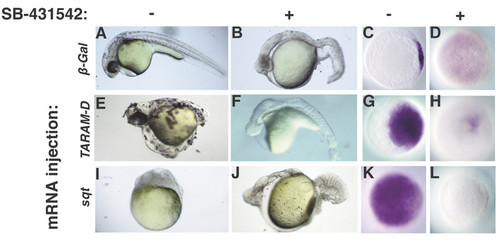
Treatment at MBT blocks the response to receptors activated during the cleavage stages. Embryos injected at the 1–4 cell stage with 10pg β-galactosidase (A-D), TARAM-D (E-H), or sqt (I-L) mRNA, and treated at 2.75 h with DMSO (A, E, I, C, G, K) or SB-431542 (B, F, J, D, H, L). (E, G) TARAM-D induces ectopic body axes and gsc expression. (F, H) The effects of TARAM-D are suppressed by treatment with SB-431542. (I, K) sqt overexpression arrests epiboly and induces ubiquitous expression of gsc. (J, L) The response to ubiquitous Sqt is blocked by treatment with SB-431542. Images of live embryos at 30 h, anterior to the left (A, B, E, F, I, J). Animal pole views of fixed embryos at 5 h, dorsal to the right (C, D, G, H, K, L). EXPRESSION / LABELING:
|

Nodal signals pattern the mesoderm during a three-hour time window. (A1-A8) Trunk somites form in embryos treated at 3.7 h (A1, 5), but flh is expressed in four ectodermal domains (A6). (B1-B8) Embryos treated at 4 h contain more somites (B5). flh is expressed at the midline and small amounts of notochord tissue are observed in live embryos (B1, arrow). pax2.1 expression is also observed (B7). At later time points, embryos have progressively more somites and more notochord (C1-E7). flh expression extends further up the midline and pax2.1 is expressed more strongly. Kupffer's vesicle forms in embryos treated 4.3 h (C3, arrowhead). shhb is not expressed in embryos treated before gastrulation (A8-E8). Images of live embryos at 24 h, anterior to the left (A1-E1; A2-E2) or 14hpf (A3-E3); dorsal views of fixed embryos at 10 h (A4-E4; A6-E6; A8-D8) or 14 h (A5-E5; A7-E7). Control embryos are depicted in Fig. 1J-S, which are from the same experiment. EXPRESSION / LABELING:
|
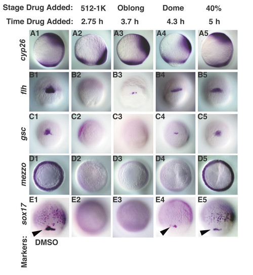
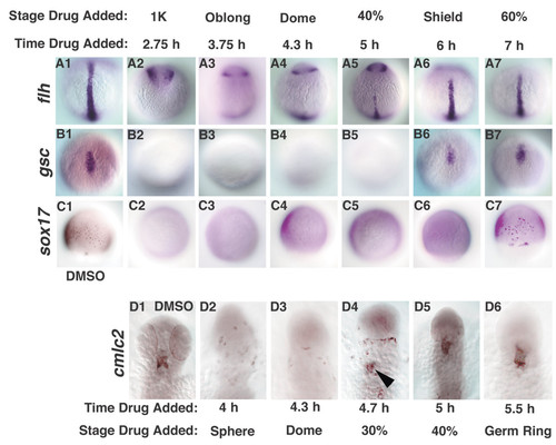
Nodal signals pattern the dorsal mesoderm and endoderm along the animal-vegetal axis in a time-dependent manner. Dorsal cell fates were examined in embryos treated with DMSO (A1-E1), or with SB-431542 at various time points. (A2-5) cyp26 expression was expressed at the margin in embryos treated at MBT, but is expressed in more animal locations at later time points. (B2-5) flh is first detected in embryos treated at 3.7 h. (C2-5) gsc is first observed in embryos treated at 4.3 h (dome stage), but is expressed at normal levels in embryos treated after 5 h (40% epiboly). mezzo transcripts are observed in embryos treated after 5 h (40% epiboly) (D5), but not at earlier stages (D2-4). sox17 is expressed in the dorsal forerunner cells in embryos treated 4.3 h (dome stage) (E4, arrowhead), but is first detected in endoderm progenitors in embryos treated at 5 h (40% epiboly) (E5). Lateral views of embryos at 10 h are depicted in A1-5, dorsal to the right. Dorsal views of embryos at 7 h (60% epiboly) (B1-C5), 5.5 h (germ ring) (D1-5) and 8 h (80% epiboly) (E1-5) are depicted. Arrowheads (E1, 4, 5) indicate sox17 in dorsal forerunner cells. All embryos are siblings. |
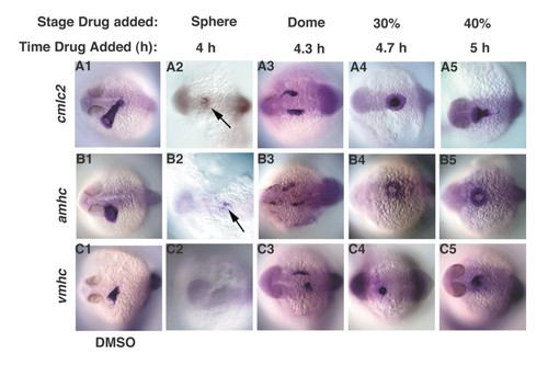
Nodal signals pattern the ventrolateral mesoderm along the animal-vegetal axis in a time-dependent manner. Heart myocardial cell fates were examined in embryos treated with DMSO (A1-C1), or with SB-431542 at various time points (A2-C5). (A2-C2) Embryos treated at 4 h, express small amounts of amhc and cmlc2, but not vmhc (arrows). (A3-3) cmlc2, amhc and vmhc are bilaterally expressed in embryos treated at 4.3 h. (A4-C5) All heart markers are expressed at the midline in embryos treated at 4.7 h. Images are dorsal views at 24 h, anterior to the left. EXPRESSION / LABELING:
|
Cell fate specification is delayed squint mutants. Cell fates were examined in sqt mutant embryos treated with DMSO (A1-D1), or with SB-431542 at various time points. (A1-7) flh was first expressed at the midline in embryos treated at 5 h (A5). (B2-7) gsc expression is first detected in embryos treated at 6 h (B6). (C2-7) sox17 expression is first detected when embryos are treated at 7 h (C7). (D1-7) cmlc2 expression was first detected in embryos treated 4.7 h (D4, arrowhead). Dorsal views of 10 h (A1-B7), 8 h (C1-C7) or 24 h (D1-D6). In D1-D6, anterior is up. The embryos in Figs. 8 and 9 are from the same clutch and weretreated in parallel, along with wild type controls (not shown). EXPRESSION / LABELING:
|
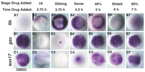
Cell fate specification is accelerated when Nodal levels are increased. Embryos were injected with sqt mRNA at the 1–4 cell stage and treated with DMSO (A1-C1) or SB-431542 at various time points (A2-C7). Sqt induces ubiquitous expression of gsc and sox17 (B1, C1) but not flh (A1). (A2-C2) SB-431542 treatment at MBT blocks expression of each of these markers. flh is strongly expressed in embryos treated at 3.7 h (A3), but fades at later time points (A4-7). gsc expression is first detected in embryos treated at 3.7 h (B3), and expands at later time points (B4-7). sox17 expressing cells are first detected when embryos are treated at 4.3 h (C4), and expands at later time points (C5-7). Animal pole views at 10 h. |